
- Usually a single firm lump 0.5–1.5 cm across (most 7–10 mm), most often on the lower leg.
- The dimple (pinch) sign — the surface dips inward when you squeeze the skin around it — is the classic clue.
- The cause is uncertain; it is sometimes linked to minor trauma such as an insect bite, but only inconsistently.
- Most are harmless and need no treatment — observation is the usual approach.
- If removed, recurrence is common (the lesion extends beyond its visible edge) and it always leaves a scar, especially on the legs.
- Freezing, shaving and laser can reduce prominence but rarely remove a dermatofibroma completely.
- Atypical or changing lesions (enlargement, ulceration, asymmetric dermoscopy) are biopsied to exclude DFSP and desmoplastic melanoma.
- Several lesions appearing together can occasionally signal altered immunity and are worth mentioning to your doctor.
A dermatofibroma is one of the most common harmless skin lumps. It is benign and does not turn into skin cancer, so most are best left alone. It usually shows up as a single firm bump — most often on the lower leg — and is frequently noticed because it catches when shaving or rubs against clothing. (1,2)
What is a dermatofibroma?
A dermatofibroma — older name cutaneous fibrous histiocytoma — is a benign nodule made of fibrous tissue (proliferating fibroblasts) in the dermis, the deeper layer of the skin. Interestingly, it is still not clear whether it is a reaction in the skin or a true growth. (1) Either way, the result is the same — a small, firm lump that tends to persist for years.
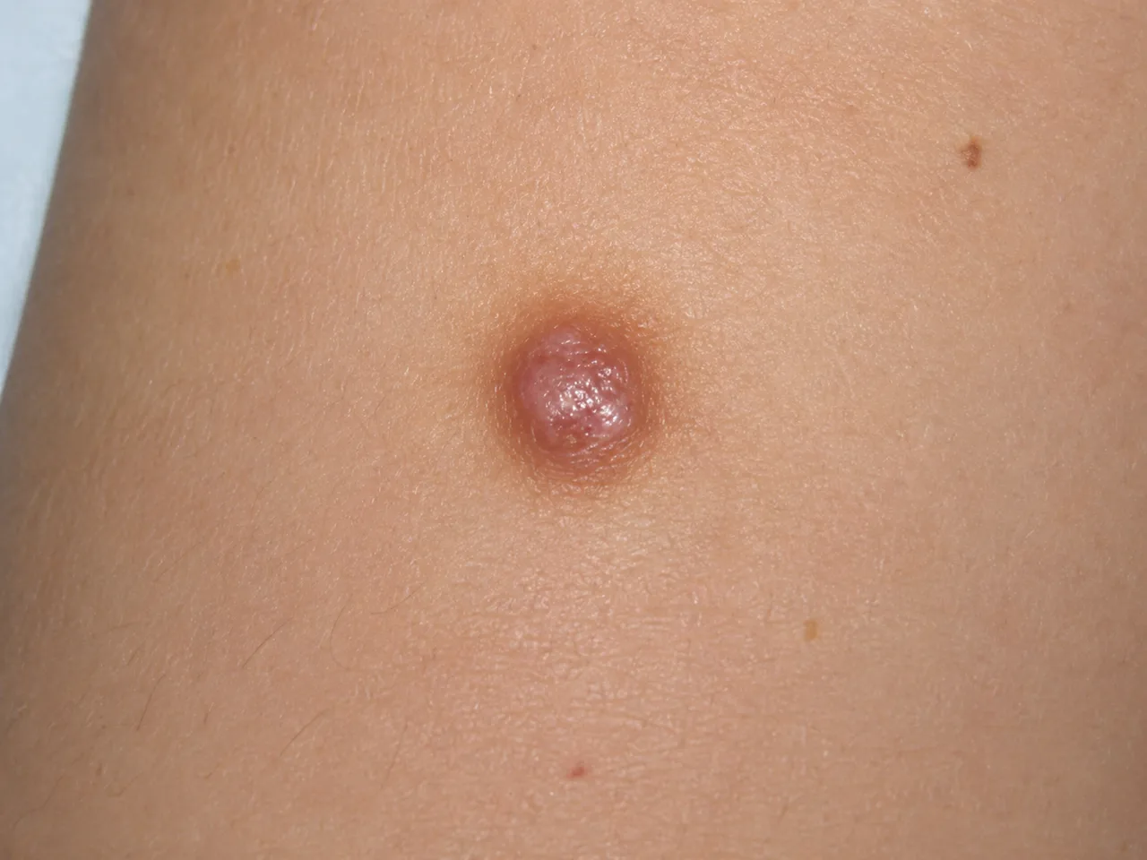
What does it look and feel like?
Typical features: (1)
- Size — usually 0.5–1.5 cm, with most around 7–10 mm
- A single firm bump, often slightly raised and dome-shaped
- Tethered to the surface but mobile over the deeper tissue — it moves with the skin rather than floating freely underneath
- Colour depends on skin tone — pink to light brown in fair skin, dark brown to black in darker skin, sometimes paler in the centre
- Usually painless, but can be painful, tender or itchy, and is easily nicked when shaving
Most people have just one. The lower leg is the classic site, but they can also appear on the arms, trunk or elsewhere. (1)
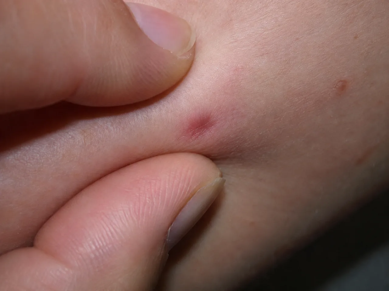
The dimple (pinch) sign
There is a simple bedside test. When you gently squeeze the skin around the lesion, its surface dimples inward instead of bulging out — the dimple (pinch) sign. (1) It happens because the lesion is anchored to the overlying skin, and it helps tell a dermatofibroma apart from other firm lumps such as cysts or lipomas, which usually do not dimple.
Why do they form?
Honestly, the cause is not fully settled. (1) Dermatofibromas are sometimes linked to minor skin trauma — an insect bite, an injection or a thorn prick — but the link is inconsistent, and plenty of lesions appear with no recognised trigger.
A few patterns are recognised: (1)
- They are more common in women than men (though some uncommon microscopic variants are seen more often in men).
- Several lesions together, or many erupting over a few months, can occasionally point to altered immunity — for example HIV, immune-suppressing medication or an autoimmune condition — and are worth mentioning to your doctor.
A dermatofibroma is not contagious and has nothing to do with hygiene.
How we diagnose it
Most dermatofibromas are recognised straight away from their appearance and feel, the dimple sign, and dermoscopy (examining the lesion under magnification). The most common dermoscopic pattern is a central white area surrounded by a faint pigment network, although this can look different in skin of colour. (1)
When a biopsy is recommended
If a lesion has an atypical feature — it has recently enlarged, ulcerated, or shows asymmetric colours and structures on dermoscopy — we recommend a biopsy or excision to confirm the diagnosis and exclude rarer look-alikes, in particular dermatofibrosarcoma protuberans (DFSP) and desmoplastic melanoma. (1) Where the microscope picture is borderline, special stains settle it.
If you are not sure what a lump is, see Other Dermatological Conditions.
Do you need treatment?
Usually not. A dermatofibroma is harmless and seldom causes symptoms, so reassurance and observation are the right answer for most people. (1)
Removal is worth considering when a lesion is:
- repeatedly nicked while shaving or catches on clothing
- tender or itchy
- changing, or
- simply bothersome to you
Removal options — what to expect
Because a dermatofibroma sits in the dermis and often extends a little beyond its visible edge, removing it completely is harder than it looks — recurrence is common even after surgical excision, and it always leaves a scar. (1) On the lower legs, where most occur, scars heal slowly and can end up more noticeable than the original lump. Freezing, shaving and laser can make a lesion less prominent but are rarely completely successful. (1)
The options below are weighed up together at consultation — and for most people, leaving a harmless lump alone is the most sensible choice.
Book a dermatofibroma assessment
If you have a firm lump you would like identified — or you are weighing up removing one — book a short review with Dr Chris Irwin. He will confirm the diagnosis (the dimple sign and dermoscopy usually make it straightforward), explain whether removal is worth it for your lesion, and plan realistically around the scar and the chance of recurrence. No referral is needed, and both clinics run the same pathway:
- Ivanhoe — Unit 1/1065 Heidelberg Road, Ivanhoe VIC 3079
- Diamond Creek — Shop 12/67 Main Hurstbridge Road, Diamond Creek VIC 3089
Symptoms
- A small, firm papule or nodule — usually 0.5–1.5 cm across, with most around 7–10 mm.
- Often slightly raised and dome-shaped; tethered to the surface skin but mobile over the deeper tissue beneath.
- Colour varies with skin tone — pink to light brown in fair skin, dark brown to black in darker skin; the centre may look paler.
- Usually a single lesion, most often on the lower leg, though they can appear on the arms, trunk or elsewhere.
- Usually painless, but can be painful, tender or itchy, and is easily nicked when shaving because it sits slightly raised.
- The classic dimple (pinch) sign — squeezing the skin around the lesion makes its surface dimple inward.
Causes & contributors
- The cause is not fully understood — it is unclear whether a dermatofibroma is a reactive process (the skin responding to something) or a true growth.
- It is made of fibrous tissue (proliferating fibroblasts) in the dermis, the deeper layer of the skin — hence the older name cutaneous fibrous histiocytoma.
- Sometimes linked to minor skin trauma — an insect bite, an injection or a thorn prick — but this association is inconsistent, and many lesions have no recognised trigger.
- More common in women than men, although some less-common microscopic variants are seen more often in men.
- Multiple dermatofibromas appearing together — or many erupting over a few months — can occasionally signal altered immunity (for example HIV, immune-suppressing medication or an autoimmune condition), and are worth mentioning to your doctor.
- Not contagious and not caused by poor hygiene.
Diagnosis
Most dermatofibromas are recognised from their appearance and feel, the dimple (pinch) sign, and dermoscopy — the most common dermoscopic pattern is a central white area surrounded by a faint pigment network, though this can look different in skin of colour. If a lesion has an atypical feature — recent enlargement, ulceration, or asymmetric colours and structures on dermoscopy — a skin biopsy or excision is recommended to confirm the diagnosis and exclude rarer look-alikes, in particular dermatofibrosarcoma protuberans (DFSP) and desmoplastic melanoma. Where the microscopic picture is in doubt, special (immunohistochemical) stains can confirm it.
Treatment options
Observation (the usual approach)
A dermatofibroma is harmless and seldom causes symptoms, so in most cases the right answer is reassurance and leaving it alone. Most are stable for years.
Surgical excision
Cutting the lesion out is the option most likely to remove it — but it leaves a linear scar, and because a dermatofibroma often extends beyond its visible edge, recurrence is common even after excision. On the lower legs, where most occur, scars also heal slowly and can end up more noticeable than the original lump. Honest counselling about scar and recurrence is essential before consenting.
Cryotherapy, shave or laser (appearance-focused) →
Freezing, shaving the surface flat, or energy-based treatments can make a lesion less prominent, but they leave the deeper part behind and are rarely completely successful — so the bump can persist or return. Best discussed alongside the other options at consultation.
When to see a doctor
See a doctor if you are unsure what a lump is and would like it confirmed, if a known dermatofibroma grows, ulcerates, bleeds or changes, if several appear together or many develop over a short time, or if a lesion is repeatedly caught when shaving or on clothing. It is also worth a review if you are weighing up removal, so the realistic scar outcome and the chance of recurrence can be planned before any procedure. Most dermatofibromas are stable and need no urgent action, but atypical or changing lesions deserve a low threshold for assessment.
Frequently asked questions
-
Is a dermatofibroma dangerous?
No. A dermatofibroma is benign and does not turn into skin cancer — it is a common, harmless growth of fibrous tissue in the dermis. The main reason for caution is that an atypical or changing lesion (one that is enlarging, ulcerating, or has asymmetric colours on dermoscopy) should be biopsied to confirm the diagnosis and exclude rarer look-alikes such as dermatofibrosarcoma protuberans (DFSP) or desmoplastic melanoma. -
What is the dimple (pinch) sign?
It is a classic bedside clue. When the skin around the lesion is gently squeezed between finger and thumb, the surface dimples inward rather than bulging out. It reflects the way the lesion is tethered to the overlying skin while staying mobile over the deeper tissue, and it helps tell a dermatofibroma apart from other firm lumps such as cysts, which usually do not dimple. -
Will it go away on its own?
Usually not. Most dermatofibromas persist for years and seldom disappear by themselves. If a lesion is not bothering you, leaving it alone is the most common and appropriate choice. -
If I have it removed, will it come back?
Often, yes. Recurrence is common even after surgical excision, because a dermatofibroma frequently extends a little beyond its visible edge. Freezing, shaving and laser are even less likely to remove it completely. This is part of why observation is usually preferred unless a lesion is genuinely bothersome — and why removal is planned around realistic expectations of both scarring and recurrence. -
Can I remove it with a cream?
No. A dermatofibroma sits in the dermis, deeper than any topical cream can reach, so creams will not remove it. For an actual dermatofibroma the options are observation or a procedure — and any product advertised as dissolving these lesions should be treated with scepticism. A lump should also be diagnosed before anything is applied to it. -
Why can the scar be worse than the original lump?
Because the lesion sits in the dermis, removing it completely means taking a deeper layer of skin and closing the wound with stitches. On the lower legs in particular — where most dermatofibromas occur — skin heals slowly and the scar can stay visible for many months. That, together with the chance of recurrence, is why we talk it through honestly before agreeing to remove one. -
What if a lump is growing or changing — could it be something else?
Occasionally. A few uncommon lesions can resemble a dermatofibroma, including dermatofibrosarcoma protuberans (DFSP) — a slow-growing, low-grade skin cancer — and desmoplastic melanoma. Features that prompt a biopsy include recent enlargement, ulceration, or asymmetric colours and structures on dermoscopy. When there is any doubt, dermoscopy plus a biopsy is the safer path. -
Should I worry if several appear at once?
It is worth mentioning to your doctor. Most people have a single dermatofibroma. When several appear together, or many erupt over a few months, it can occasionally reflect altered immunity — for example HIV, immune-suppressing medication or an autoimmune condition — so it is reasonable to review the wider picture. -
Do I need a referral?
No referral is required — you can book directly. A referral from your GP or other treating clinician is welcome (it helps with continuity of care) but is not a barrier to being seen. Both clinics (Ivanhoe and Diamond Creek) operate the same pathway.
References
Related
Related treatments
Medically reviewed by Dr Christopher Irwin, MBChB, FRACGP, MMed (Skin Cancer), FACAM, MSCCA · Published 2026-06-06 · Updated 2026-06-30 · Editorial policy