
- Dysplastic naevus is a histological diagnosis made by a pathologist — not a clinical appearance alone.
- Most dysplastic naevi never become melanoma; they are markers of increased risk rather than direct precursors.
- The 2018 WHO classification uses a two-tier grading system — low-grade and high-grade.
- Low-grade dysplastic naevi are typically managed with a 2–3 mm excision margin.
- High-grade dysplastic naevi are managed like melanoma in situ — typically with a ~5 mm re-excision margin.
- Most melanomas (~70–80%) arise on previously normal skin, not from existing moles — whole-body surveillance matters more than watching only the dysplastic lesion.
- Photographic and dermatoscopic monitoring is preferred over removing every atypical-looking mole.
- Patients with multiple dysplastic naevi need regular skin checks, monthly self-examination and strict sun protection.
- FAMMM (Familial Atypical Multiple Mole Melanoma) syndrome implies significantly higher lifetime melanoma risk and first-degree-relative surveillance.
- The "ugly duckling" — the mole that looks different from the patient's other moles — is the single most useful self-examination signal.
Quick definition
A dysplastic naevus is a type of mole that is benign (non-cancerous) but shows architectural disorder and cellular atypia under the microscope. It is a histological diagnosis — confirmed by a pathologist on a tissue sample, not by the way the mole looks on the skin. Most dysplastic naevi never become melanoma. They are best understood as a marker of increased melanoma risk rather than a direct precursor. The 2018 WHO classification uses two grades — low-grade (re-excised with 2–3 mm of normal skin) and high-grade (re-excised with about 5 mm, the same margin used for melanoma in situ). For patients with multiple atypical moles, photographic and dermatoscopic monitoring is preferred over removing every lesion.
What is a Dysplastic Naevus?
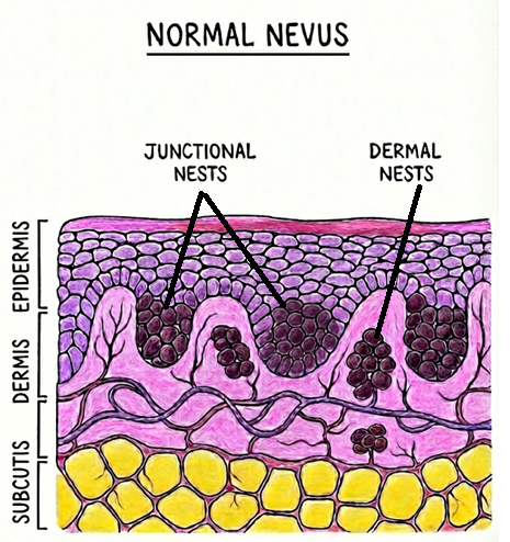
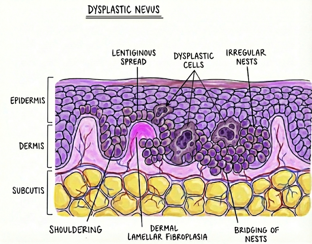
A dysplastic naevus is a type of mole that appears benign (non-cancerous) but has some unusual features when examined under a microscope. The term comes from pathology: “dysplastic” means the cells look atypical or slightly abnormal (but not cancerous) in their arrangement or appearance, and “naevus” means a mole (a growth made of pigment-producing melanocyte cells). In other words, a dysplastic naevus is a mole where the microscopic structure is not entirely normal. Importantly, this is a histological diagnosis — it is determined by a pathologist looking at a tissue sample under the microscope, rather than by appearance alone on the skin.
-
How it differs from a normal mole. Under the microscope, a dysplastic naevus shows an architectural disorder — the way the melanocyte cells are arranged in the skin is somewhat disorganised compared to a common mole. The nests of melanocytes may spread out sideways beyond the main centre of the mole (called “shouldering” or lateral extension) and might form bridges between neighbouring rete ridges. There is often a thin band of fibrous tissue (lamellar fibrosis) underlying the mole and a mild inflammatory response around it. The cells also show atypia, meaning the nuclei are a bit larger, irregular or darker than usual. These changes are less pronounced than what is seen in melanoma, but more than in an ordinary mole.
-
How it differs from an “atypical mole” diagnosed by sight. Many dysplastic naevi look perfectly ordinary on the skin. In fact, many moles that are found to be “dysplastic” under the microscope can look completely normal on the skin. The diagnosis of a dysplastic naevus really comes from the microscope examination — not from appearance alone.
What Causes Dysplastic Naevi and Who Gets Them?
Dysplastic naevi are thought to develop due to a mix of genetic factors and environmental influences:
-
Genetics. Some people inherit a tendency to have multiple dysplastic moles. There is a familial condition (sometimes called atypical mole syndrome or FAMMM — Familial Atypical Multiple Mole Melanoma syndrome) where individuals have dozens of dysplastic naevi and a family history of melanoma. However, you do not need to have a family syndrome to develop dysplastic naevi.
-
Sun exposure. UV light from the sun (or tanning beds) is a known contributor. Episodes of intense sun exposure or sunburns, especially in childhood, may increase the likelihood of developing atypical moles. Dysplastic naevi often appear in sun-exposed areas (like the back), but they can also occur in sun-protected areas, suggesting that sun is only part of the story.
-
Skin type. People with fair skin, light hair and eye colour, and those who freckle easily tend to develop more moles (including atypical ones). However, dysplastic naevi can occur in anyone.
Are Dysplastic Naevi Precancerous?
This is an important and sometimes confusing question. The truth lies somewhere in between and is still being studied:
-
Not exactly “pre-cancer” in the way actinic keratoses are. An actinic keratosis is a sun-damaged spot that can turn into a squamous cell carcinoma if not treated. Dysplastic naevi are not like that — the majority of dysplastic moles do not ever turn into melanoma. In fact, most melanomas (about 70–80%) seem to arise de novo (on previously normal skin) rather than from an existing mole.
-
Some may represent early changes toward melanoma. Particularly when a dysplastic naevus has high-grade atypia, the line between a dysplastic naevus and an early melanoma in situ can be very fine. Pathologists sometimes even disagree — one might call it “severely dysplastic naevus” while another might call the same sample “early melanoma.”
- A marker of risk (red flag) for melanoma. The most accepted view today is that a histologically dysplastic naevus is more of a risk marker than a direct precursor. In other words, having these atypical moles on your skin indicates that your skin is the type that is prone to melanoma. The dysplastic naevus itself might never become cancerous, but the presence of dysplastic naevi increases the chance that somewhere on your skin, a melanoma could develop over time.
Do Dysplastic Naevi Increase Melanoma Risk?
Yes. Having histologically confirmed dysplastic naevi is associated with a higher risk of melanoma developing in the future, and often that melanoma is an entirely new lesion — not the mole that was dysplastic, but elsewhere on the skin.
-
Overall risk. Studies have shown that people with multiple dysplastic naevi have a significantly elevated risk of melanoma. Having more than five dysplastic moles may correspond to about a 10-fold increase in melanoma risk relative to someone with none. The Skin Cancer Foundation notes that individuals with 10 or more atypical moles might have roughly 12 times the risk of melanoma.
-
Melanomas arising from dysplastic naevi. Experts estimate that about 20–30% of melanomas may develop in association with an atypical (dysplastic) mole. This still means the majority of melanomas do not come from existing moles. So if you have had a dysplastic mole removed, it does not mean you are “safe” from melanoma in the future — you still need regular skin checks of your entire skin.
-
Family history and syndrome. If dysplastic naevi run in your family and especially if combined with a family history of melanoma, the risk is even higher. People with dysplastic naevus syndrome (very large number of moles and a strong family incidence of melanoma) have a lifetime melanoma risk of 50% or higher.
Grading: Low-Grade vs High-Grade
Pathologists grade the degree of dysplasia (atypia) present. The 2018 WHO Classification of Skin Tumours simplified the system into two tiers:
-
Low-grade dysplastic naevus. Corresponds to the lesser degrees of atypia. What might have been called “mildly” or “moderately” dysplastic before would now be termed low-grade. These have only mild to moderate cell atypia under the microscope.
-
High-grade dysplastic naevus. Corresponds to what pathologists used to call severely dysplastic naevus. The cells are quite atypical — often approaching the appearance of melanoma in situ. In fact, a high-grade dysplastic naevus can be so atypical that some experts consider it essentially the same as a melanoma in situ.
The reason for this change is that the moderate category was somewhat subjective and pathologists did not always agree on what was moderate versus mild or severe. By simplifying it, the hope is to clarify management.
Treatment and Management
The good news is that dysplastic naevi can be cured by complete removal — they do not “spread” if fully excised with a clear margin. The main question is how wide a margin is needed.
-
Complete excision is the standard for any confirmed dysplastic naevus. For a low-grade dysplastic naevus, a safety margin of about 2–3 mm of normal skin around the mole is considered sufficient.
-
High-grade dysplastic naevus management. Because a high-grade dysplastic naevus can look so much like melanoma in situ under the microscope, the treatment approach is similar to melanoma in situ — typically a re-excision with about a 5 mm margin of normal skin.
-
Monitoring and follow-up. Removing a dysplastic naevus takes care of that mole, but because these moles are markers of risk, your doctor will likely recommend regular skin checks after that. Typically, if you have had one or a few low-grade dysplastic naevi, an annual full skin examination by a skin cancer doctor is advised — or more often if you have many of them or other risk factors. You should also perform monthly self-skin exams at home, keeping an eye on any changing moles or new lesions.
-
Sun protection. Diligent use of broad-spectrum sunscreen (SPF 50 or higher), wearing protective clothing and hats, and avoiding indoor tanning are all strongly recommended.
References
- National Cancer Institute. Common Moles, Dysplastic Nevi, and Risk of Melanoma.
- Menzinger S et al. Dysplastic Nevi and Superficial Borderline Atypical Melanocytic Lesions. Dermatopathology. 2025.
- Skin Cancer Foundation. Atypical Moles & Your Skin. 2022.
- Calabresi L. Dysplastic naevi: the controversy continues. Healthed Clinical Articles. 2019.
- WHO Classification of Skin Tumours, 4th edition. 2018.
Symptoms
- Many dysplastic naevi look like ordinary moles on the skin — diagnosis is histological, not visual.
- Some are clinically atypical — asymmetric, irregular border, varied colours (tan, brown, pink, black), larger than 6 mm.
- May show "fried egg" appearance — a darker central area within a paler surrounding flat zone.
- Often arise in adolescence or early adulthood and may continue to develop into middle age.
- Frequently multiple — a single isolated dysplastic naevus is less common than several across the back, chest or limbs.
- A "different-looking" mole — the ugly duckling sign — stands out from the patient's other moles and warrants attention.
Causes & contributors
- Genetic predisposition — inherited tendency for atypical moles is common, particularly with fair skin and many naevi.
- Familial Atypical Multiple Mole Melanoma syndrome (FAMMM) — multiple dysplastic naevi plus family history of melanoma; significant lifetime melanoma risk.
- UV exposure — intense sun exposure and sunburns (especially in childhood) contribute, including from tanning beds.
- Fair skin, light hair, light eye colour, freckle tendency (Fitzpatrick I–II) — higher background mole burden including atypical moles.
- Sun-protected sites also affected — dysplastic naevi can develop on the buttocks, scalp and other rarely-exposed sites, so UV is contributor but not the whole story.
Diagnosis
Dysplastic naevus is a histological diagnosis — confirmed by a pathologist on a tissue sample after biopsy or excision, not by clinical appearance alone. Many moles that look ordinary on the skin turn out to be dysplastic under the microscope, and conversely many clinically atypical-looking moles are not histologically dysplastic. At The Skin Doctor, lesions of clinical or dermatoscopic concern are removed in full (rather than partially biopsied where feasible) and sent for histology, with grade — low or high — confirmed in the pathology report. Where lesions are very numerous, photographic and dermatoscopic monitoring at planned intervals is used to identify the lesion most worth removing for histology rather than removing them all.
Treatment options
Watchful waiting with photographic monitoring
For clinically benign-looking lesions in a patient with multiple naevi, planned-interval photographic and dermatoscopic review identifies changing lesions for histology rather than removing every mole. Self-examination at monthly intervals between visits is part of this pathway.
Diagnostic excision (clinical concern)
Lesions of clinical or dermatoscopic concern are removed in full and sent for histology. This is preferred over partial biopsy where feasible because a partial sample can miss the most atypical area.
Re-excision for low-grade dysplastic naevus
A safety margin of about 2–3 mm of normal skin is typically considered sufficient for histologically confirmed low-grade dysplasia. Recurrence after clear margins is uncommon and routine wider re-excision is generally not required.
Re-excision for high-grade dysplastic naevus
Because high-grade dysplastic naevus is histologically close to melanoma in situ, the standard re-excision margin is about 5 mm of normal skin — the same margin used for melanoma in situ.
Long-term skin-check and self-examination follow-up
Because dysplastic naevi are markers of risk rather than direct precursors, the most important treatment is long-term surveillance — annual or more frequent full-body skin checks by a skin-cancer doctor, monthly self-examination, and prompt review of any changing or new lesion.
Strict daily sun protection
SPF 50+ broad-spectrum sunscreen daily, protective clothing, hats, sunglasses, avoidance of intense midday UV and complete avoidance of tanning beds. Sun protection does not eliminate risk but is the most modifiable component of melanoma prevention.
When to see a doctor
Book a doctor review if you have any of the following: a new or changing mole (size, colour, shape, bleeding, itching); a mole that looks different from your other moles (the "ugly duckling"); multiple atypical-looking moles, particularly with a personal or family history of melanoma; a previously diagnosed dysplastic naevus that you would like included in a structured monitoring program; or fair skin and significant childhood sun exposure with concerns about background risk. A new pigmented lesion appearing after age 40 is always worth assessing. Patients with five or more atypical-looking moles, especially with a family history of melanoma, should establish regular full-body skin-check surveillance.
Frequently asked questions
-
What does it mean if a mole is called dysplastic?
It means the mole has atypical features under the microscope — the way the melanocyte cells are arranged is somewhat disorganised compared to a common mole, and the cells themselves look slightly more abnormal than usual. It does not mean the mole is cancer. Dysplastic naevus is a histological diagnosis made by a pathologist on a tissue sample — not a diagnosis made just by looking at the mole on the skin. Many dysplastic naevi look perfectly ordinary on the skin. -
Is a dysplastic naevus the same as melanoma?
No. A dysplastic naevus is a benign mole with some atypical features — not cancer. Most dysplastic naevi never become melanoma. However, high-grade dysplastic naevi can look very similar to early melanoma (melanoma in situ) under the microscope, and the line between them can be thin enough that experienced pathologists sometimes disagree. This is why high-grade dysplastic naevi are managed with the same wider re-excision margin used for melanoma in situ. -
Do I need to have all my moles removed if I have dysplastic naevi?
No — and we generally recommend against it. Removing every mole is impractical and unnecessary. The right approach for patients with multiple naevi is photographic and dermatoscopic monitoring at planned intervals, with selective excision of any lesion that is changing or genuinely atypical. The goal is to identify the few lesions worth removing for histology, not to remove them all. Most dysplastic naevi remain dysplastic and never progress. -
What's the difference between a low-grade and a high-grade dysplastic naevus?
The 2018 WHO classification uses a two-tier system. Low-grade corresponds to what was previously called mildly or moderately dysplastic — only mild-to-moderate atypia under the microscope, managed with a 2–3 mm re-excision margin. High-grade corresponds to severely dysplastic naevus — cells approach the appearance of melanoma in situ, and the lesion is managed with the same ~5 mm re-excision margin used for melanoma in situ. The grade is determined by the pathologist on the tissue sample, not visually. -
How wide a margin is needed to remove a dysplastic naevus?
It depends on the histological grade, not on the clinical appearance. Low-grade dysplastic naevus — typically 2–3 mm of normal skin around the lesion. High-grade dysplastic naevus — typically about 5 mm, the same margin used for melanoma in situ. For lesions of initial clinical concern, the first excision is diagnostic (in full, with a small margin) and is followed by a definitive re-excision once histology confirms grade. -
Will I get melanoma if I have dysplastic naevi?
Not necessarily — but your risk is higher than average. Having more than five dysplastic naevi is associated with roughly a 10-fold increase in melanoma risk relative to someone with none. Ten or more atypical-looking moles may correspond to approximately 12 times the average risk. However, most melanomas (about 70–80%) arise on previously normal skin — not from existing moles. This is why whole-body surveillance matters more than just watching the dysplastic lesions themselves. -
How often should I have skin checks if I have dysplastic naevi?
Most patients with one or a few low-grade dysplastic naevi are reviewed with an annual full-body skin check. Patients with multiple atypical moles, prior melanoma, FAMMM syndrome or strong family history of melanoma may be reviewed more frequently — every 3–6 months in some cases. Between visits, monthly self-examination at home is recommended, looking for changing moles or new lesions. The exact interval is individualised at consultation based on your risk profile. -
Are my children at risk if I have dysplastic naevi?
There can be a familial tendency to develop dysplastic naevi, and in FAMMM syndrome (multiple dysplastic naevi plus family history of melanoma) the risk to first-degree relatives is substantial. If you have a strong personal and family history, first-degree relatives benefit from establishing skin-check surveillance and learning self-examination. UV protection from childhood — particularly avoiding sunburns — is the most modifiable factor. This is discussed at consultation where relevant.
References
- Dysplastic nevus part I — historical perspective, classification, and epidemiology. J Am Acad Dermatol. 2023.DOI: 10.1016/j.jaad.2022.04.068
- Dysplastic nevus part II — molecular/genetic profiles and management. J Am Acad Dermatol. 2023.DOI: 10.1016/j.jaad.2022.05.071
- Risk of subsequent cutaneous melanoma in moderately dysplastic nevi excisionally biopsied but with positive histologic margins. JAMA Dermatol. 2018.DOI: 10.1001/jamadermatol.2018.3359
Related
Medically reviewed by Dr Christopher Irwin, MBChB, FRACGP, MMed (Skin Cancer), FACAM, MSCCA · Last reviewed 2026-06-06 · Editorial policy