
- Melanoma in situ (Stage 0) is the earliest possible melanoma stage — confined to the epidermis only, above the basement membrane.
- There is no route to metastasis because the vessels cancer uses to spread (blood, lymph) exist only in the dermis, not in the epidermis.
- Treatment is simple surgical excision with appropriate margins — that is the only treatment required.
- No CT, PET or lymph node biopsy is indicated; the absence of testing reflects the excellent prognosis.
- Follow-up is regular skin checks — Dr Chris reviews patients every 3 months for the first two years, then extends to 6–12 monthly.
- Patients with one prior melanoma have a 9× higher risk of a second primary melanoma (Bradford et al, Arch Dermatol 2010); risk is highest in the first two years.
- Scar care options include silicone gel (Strataderm, twice daily for 3 months) and MEFIX breathable dressing (6–12 weeks).
- Care provided across two Melbourne clinics — Ivanhoe and Diamond Creek.
Quick definition
Melanoma in situ (also called Stage 0 melanoma) is the earliest possible stage of melanoma. The cancer cells are confined to the epidermis — the top layer of skin — and have not crossed the basement membrane into the dermis below. Because the blood and lymph vessels cancer uses to spread exist only in the dermis, melanoma in situ has no route to metastasis and is essentially always cured by simple surgical excision. No CT, PET or lymph node biopsy is required.
Being diagnosed with melanoma can be scary, but the good news is that the type of melanoma you have is very early-stage and can be completely cured. This handout explains melanoma in situ — and why it does not require additional tests like lymph node biopsies or body scans. We’ll also discuss what to expect next and how these early melanomas are managed in a patient-friendly way.
Melanoma In Situ (Stage 0)
Melanoma is a cancer of pigment cells (melanocytes) in the skin.
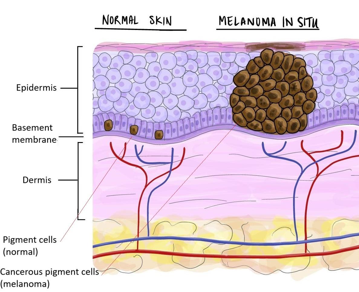
Melanoma in situ means the cancer cells are confined to the very top layer of the skin (the epidermis). It is sometimes called Stage 0 melanoma. In plain language, it means the melanoma “hasn’t started digging deeper into the skin” yet. The “in situ” part just means that it is still “in place” — ie. that it is still in the epidermis only and hasn’t learnt to invade yet.
The skin contains two basic parts. The epidermis is the top layer of skin and the bottom layer of skin is called the dermis. The epidermis and dermis are separated by a very tough canvas-like layer called the basement membrane or “dermo-epidermal junction”. If a cancer has not yet learnt how to get through this tough canvas layer, that means it has no way of travelling to other parts of the body (metastasis). This is because all of the highways that cancers use (eg. veins, arteries and lymph vessels) are all only in the dermis. There are no highways in the epidermis. This is great because it means if we cut it out properly with good margins, the cancer can essentially be cured with no risk of metastasis and very low risk of recurrence.
No lymph node biopsy or scans needed
Since it cannot spread to lymph nodes or organs (it hadn’t reached those deeper channels), no further tests like lymph node biopsies, CT scans, or PET scans are needed. Your doctors know the melanoma was only on the surface, so surgery is the only treatment necessary.
Follow-up
The main follow-up for melanoma in situ is regular skin checks. Every doctor’s practice is different. We know that a patient who has been diagnosed with one melanoma has a 9× greater risk of developing a second melanoma compared to a matched control (Bradford PT, Freedman DM, Goldstein AM, Tucker MA. Increased risk of second primary cancers after a diagnosis of melanoma. Arch Dermatol. 2010;146(3):265-272.) and this is greatest in the first two years after diagnosis.
- No routine scans or blood tests. As discussed, you won’t be undergoing regular CT/PET scans or blood work for melanoma markers, because there’s no indication to do so. Don’t be alarmed by the lack of testing — it truly is a positive sign about your prognosis.
- Scar care. You will have a scar from the surgery. Keep it clean and protected from the sun as it heals. Over time (months to a year) it will fade and soften.
You can help minimise the scarring by using a silicone gel called strataderm twice a day for 3 months. We will chat more about the specific restrictions for your specific wound.
Another technique to reduce scarring is to keep the wound dressed for as long as possible after surgery. This sounds like a lot more effort than it is — after the initial sutures come out our nurses will show you how to dress the wound with MEFIX. This is a breathable dressing that you can get wet, pat dry and essentially just treat as a second skin. What this dressing does is every time you move the area over the next 6–12 weeks, the dressing takes some of the stretch instead of making the scar stretch a little. You change the MEFIX every 5–7 days depending on activity level, soak it off with a little bit of olive oil (or just in the shower) and gently peel it off, before drying the skin and applying another MEFIX dressing. The longer you do this for the better the wound will be. In general for facial wounds we recommend the silicone gel due to its ease of use.
Scarring is inevitable but I try my absolute best to give you the best scar possible using the best suturing techniques.
- Sun protection. One of the best ways to prevent future skin cancers is to practice good sun safety. That means using sunscreen (SPF 50) on exposed skin daily, wearing hats and protective clothing when in strong sun, and avoiding indoor tanning.
- Peace of mind. It’s normal to feel anxious after a cancer diagnosis, even one as low-risk as this. Remember that your melanoma was caught at a very early stage. If you find yourself worrying a lot, consider discussing these feelings with your doctor. Sometimes connecting with a counsellor or a support group of melanoma survivors can be helpful. Knowing others have gone through the same thing and are doing fine can be very reassuring.
Frequently asked questions
-
Is melanoma in situ cancer?
Yes, technically — but it is the earliest possible stage (Stage 0) and is essentially always curable. The cancer cells are still confined to the very top layer of skin (the epidermis), above the basement membrane that separates epidermis from dermis. Because the deeper vessels (blood and lymph) only exist in the dermis, the tumour has no route to spread. -
Can melanoma in situ spread to lymph nodes or other organs?
No. By definition, melanoma in situ has not crossed the basement membrane into the dermis, and the vessels that cancer uses to travel only exist in the dermis. There are no "highways" in the epidermis — so no route to metastasis. -
Do I need a CT scan, PET scan or lymph node biopsy?
No. Since melanoma in situ cannot spread, no further imaging or lymph node biopsy is indicated. The lack of testing is a positive sign about your prognosis, not an oversight. -
How is melanoma in situ treated?
Surgical excision with appropriate margins is the only treatment needed. The piece of skin containing the melanoma (plus a margin of normal-looking surrounding skin) is removed and sent to a pathologist to confirm clear edges. -
How often should I have skin checks after a melanoma in situ?
Dr Chris generally reviews patients every 3 months for the first two years (when the risk of a second primary is highest), then gradually extends to every 6–12 months on a case-by-case basis. Patients with one prior melanoma have a 9× higher risk of a second primary melanoma (Bradford et al, Arch Dermatol 2010). -
How can I minimise the scar?
Two evidence-based options. (1) Silicone gel (e.g. Strataderm) applied twice daily for 3 months — best for facial wounds because it is easy to use. (2) MEFIX dressing — a breathable, water-resistant tape worn for 6–12 weeks and changed every 5–7 days. The dressing takes some of the movement-related stretch off the healing wound, which can meaningfully improve the final scar. -
Will I get another melanoma?
Your risk of a second primary melanoma is about 9× higher than the general population (Bradford et al, Arch Dermatol 2010), and that risk is highest in the first two years. This is why structured surveillance and lifelong sun protection matter — not because the original is likely to come back, but because new ones can appear on other parts of the body.
References
- Clinical practice guidelines for the diagnosis and management of melanoma. Cancer Council Australia & Melanoma Institute Australia.
- Melanoma in situ — Part I. Epidemiology, screening, and clinical features. J Am Acad Dermatol. 2015.DOI: 10.1016/j.jaad.2015.04.014
- Cutaneous melanoma. Lancet. 2023. (Long GV, Swetter SM, Menzies AM, Gershenwald JE, Scolyer RA.)DOI: 10.1016/S0140-6736(23)00821-8
Related
Medically reviewed by Dr Christopher Irwin, MBChB, FRACGP, MMed (Skin Cancer), FACAM, MSCCA · Last reviewed 2026-06-06 · Editorial policy