
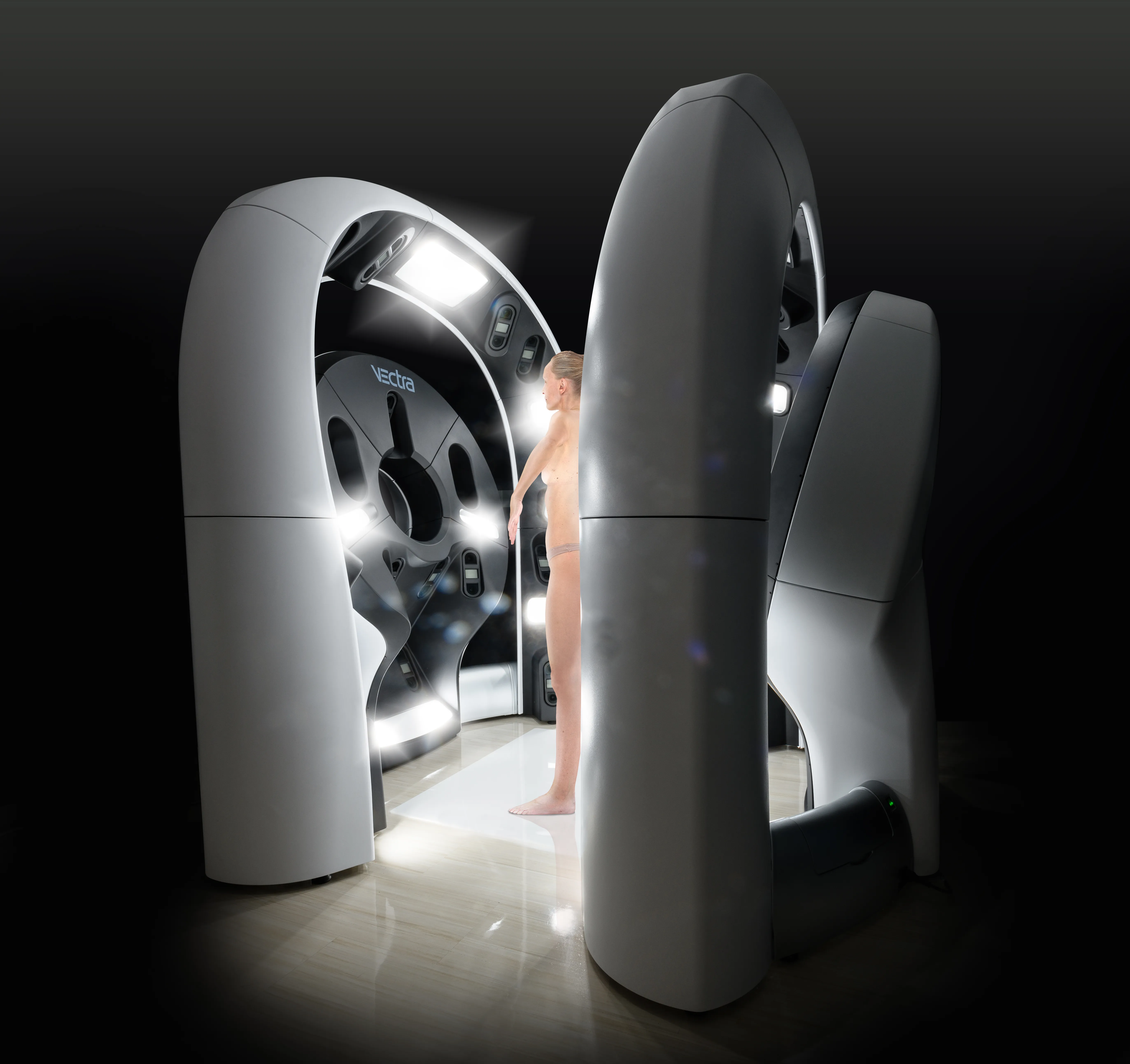
- 92 calibrated digital cameras capture your entire skin surface in a single moment.
- 3D avatar can be rotated and zoomed from any angle for detailed review.
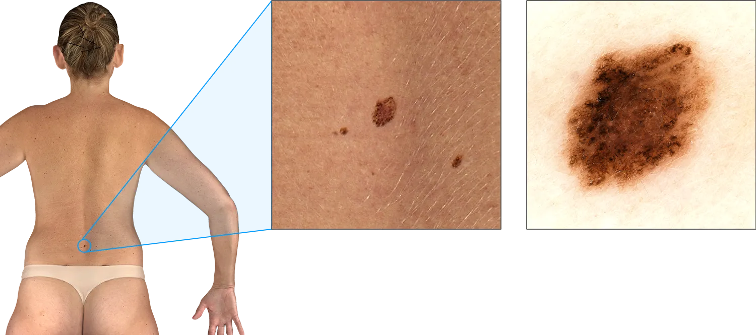
- Each lesion can be virtually "lifted" from the skin for inspection and measurement.
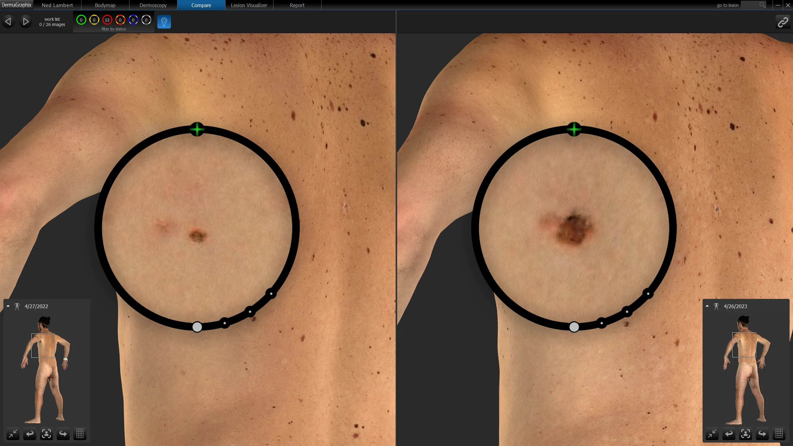
- Powerful change detection between scans under standardised conditions.
- Particularly valuable for patients with many moles or a melanoma history.
- Also used for conditions tracked by surface area — psoriasis, vitiligo, sun damage.
VECTRA® WB360 3D Imaging
World-first 3D total-body imaging at our Diamond Creek clinic — every spot mapped, time-stamped, tracked.
At our Diamond Creek clinic, the VECTRA® WB360 captures a complete, high-resolution digital record of your skin in just a few seconds. Purpose-built for dermatology, it reconstructs a lifelike 3D avatar of your body, mapping every visible mole and lesion from head to toe. This isn’t a “photo booth” — it is a specialised medical imaging platform that underpins some of the most advanced skin-cancer surveillance programs in the world.
Why 3D mole mapping matters
Traditional 2D body photography has helped monitor moles for years, but it can miss areas on curved or hard-to-see surfaces and often needs many separate images. The VECTRA® WB360 changes that.
Complete coverage in a single capture
The system images almost your entire skin surface at once — including the sides of the body and curved areas that conventional photography struggles with — and reconstructs it as a 3D avatar you and your doctor can rotate and zoom from any angle.

A precise location for every lesion
Each spot is given an exact position on your 3D map, so lesions can be reliably relocated at future visits — nothing is forgotten or “lost” between appointments. Individual lesions can be virtually “lifted” from the skin and inspected in close-up, with dermoscopic images linked to the right point on the map.
Powerful change detection over time
Because your whole skin is captured under standardised conditions, repeat scans can be compared side by side to highlight new lesions and subtle changes in existing moles that might otherwise be overlooked.
Ideal for patients with many moles — and beyond
Some patients have hundreds or even thousands of pigmented lesions, and a 3D system is especially valuable for tracking complex patterns over time. VECTRA is also used internationally to document conditions measured by surface area — psoriasis, vitiligo and extensive sun damage — calculating affected areas and showing how they change.
What to expect
-
Step into the VECTRA® WB360 booth
Inside the unit are 92 digital cameras arranged in a circular frame around you.
-
All cameras fire at once
In a single moment, the system captures your entire exposed skin surface in macro-quality resolution. No multiple poses, no dozens of separate photos — one fast, standardised capture.
-
3D avatar reconstruction
Specialised software stitches the images together to reconstruct a high-resolution 3D model of your body. Every freckle, mole and mark is mapped onto this avatar, which can be rotated and zoomed from any angle.
-
Lesion-level analysis
Each pigmented lesion can be virtually "lifted" from the skin on the 3D model so clinicians can inspect and measure it in detail. Where needed, dermoscopic images are linked to the relevant point on your map.
-
Secure storage and comparison
Your 3D images are stored on a secure server. Future scans can be compared side-by-side with your previous avatar, helping detect subtle changes or new lesions across your entire skin surface.
Results timeline
- Same appointment 3D avatar reviewed by your doctor as part of the full skin examination.
- 2–3 years (typical) VECTRA® WB360 scan repeated; 2D imaging and clinical examination performed more frequently in between.
- Indefinite Securely stored images allow lifelong lesion comparison across visits.
Ideal candidate
- Patients having a full skin check at our Diamond Creek clinic.
- People with many moles — hundreds or even thousands of pigmented lesions.
- Patients with a previous melanoma or strong family history of melanoma.
- Anyone wanting the most advanced surveillance technology available.
- Patients with conditions documented by surface-area change (psoriasis, vitiligo, extensive sun damage).
Frequently asked questions
-
Is this just a photo booth?
No. The VECTRA® WB360 is a highly specialised medical imaging platform supporting some of the most advanced skin cancer surveillance programs in the world. -
Why 3D rather than 2D mapping?
2D photography can miss areas on curved or hard-to-see surfaces. VECTRA® WB360 captures almost your entire skin surface in a single capture, including sides of the body and curved areas, and gives each spot a precise 3D location. -
Is it safe?
Yes. There is no radiation, no contact and no discomfort — it is purely photography from 92 calibrated cameras. -
Can VECTRA help with conditions other than skin cancer?
Yes. VECTRA 3D is used internationally to document and monitor conditions such as psoriasis, vitiligo and extensive sun damage, because it can calculate affected surface areas and show how they change over time.
References
- Total body photography for the diagnosis of cutaneous melanoma in adults — a systematic review and meta-analysis. Br J Dermatol. 2021.DOI: 10.1111/bjd.19759
- The value of total body photography for the early detection of melanoma — a systematic review. Int J Environ Res Public Health. 2021.DOI: 10.3390/ijerph18041726
Related
Medically reviewed by Dr Christopher Irwin, MBChB, FRACGP, MMed (Skin Cancer), FACAM, MSCCA · Last reviewed 2026-06-06 · Editorial policy