
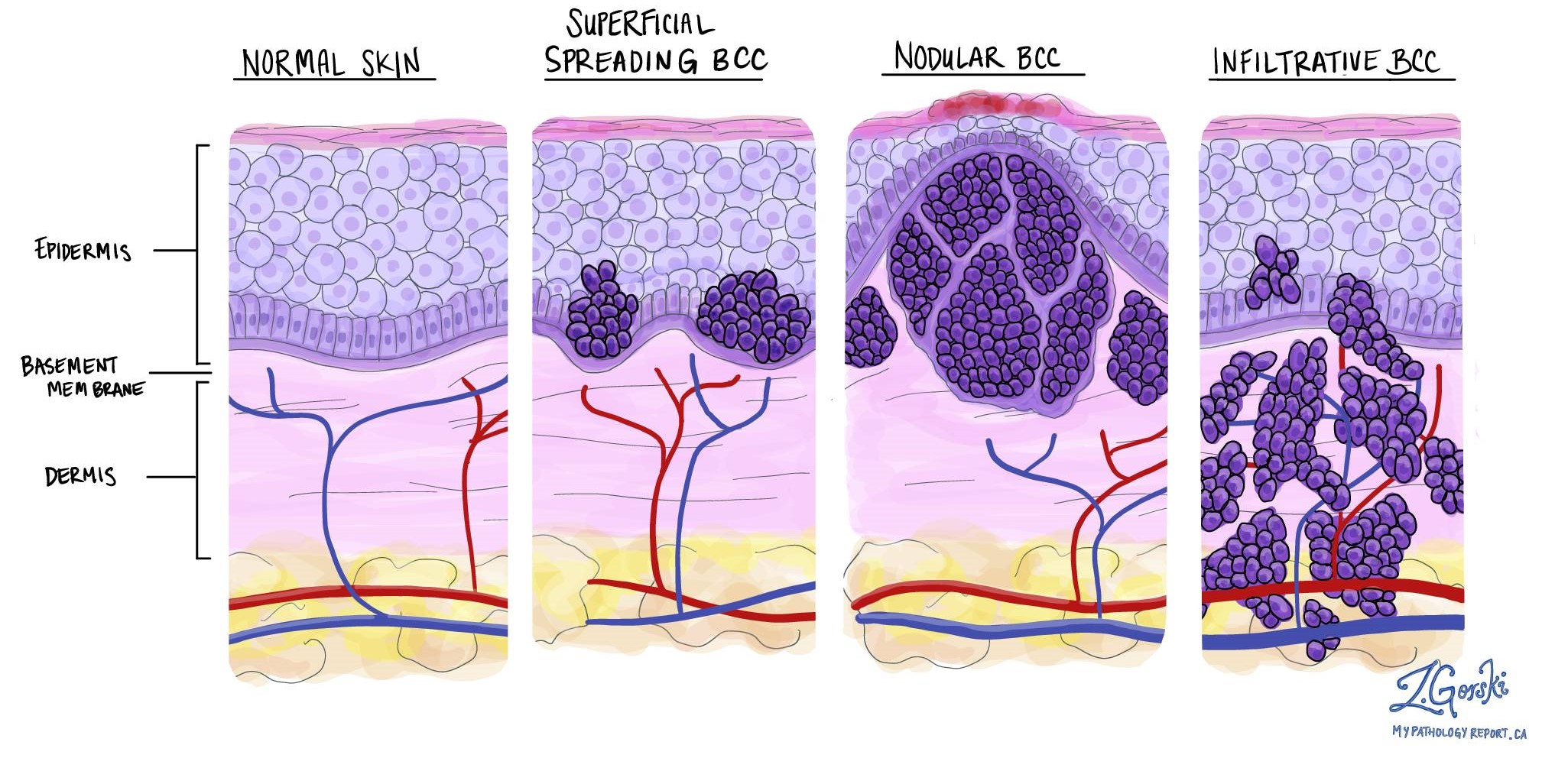
- Invasive BCC has grown through the basement membrane into the dermis — but "invasive" refers to skin depth only, not body-wide spread.
- Nodular BCC (~60% of all BCCs) is the most common subtype — a shiny pearly nodule, often on the face, with tiny visible blood vessels (telangiectasias).
- Infiltrative and morpheaform (sclerosing) BCCs (5–10% of BCCs) look like flat scar-like patches with indistinct edges and need wider surgical margins.
- "Rodent ulcer" is an older term for a nodular BCC with a central ulcer or crater.
- Even invasive BCCs metastasise in less than 0.1% of cases — routine CT/MRI scans, blood tests and lymph node biopsy are not indicated.
- Surgical excision with a 3–5 mm margin is first-line — cure rate 95–98%.
- Mohs micrographic surgery is reserved for selected high-risk sites (face — nose, eyelids, ears, lips) or recurrent BCCs.
- About 50% of patients develop another non-melanoma skin cancer within 5 years — structured skin checks and sun protection matter.
- Care provided across two Melbourne clinics — Ivanhoe and Diamond Creek.
Quick definition
Invasive basal cell carcinoma is a BCC that has grown through the basement membrane into the dermis — the layer below the epidermis. The word “invasive” here refers to depth in the skin only, not body-wide spread. Even invasive BCCs metastasise in less than 0.1% of cases. The main subtypes are nodular (~60% of all BCCs — a pearly bump often on the face), infiltrative, and morpheaform/sclerosing (5–10%, scar-like patches with indistinct edges). First-line treatment is surgical excision with a 3–5 mm margin — cure rate 95–98% for primary tumours. Mohs surgery is reserved for selected high-risk sites (face — nose, eyelids, ears, lips) or recurrent BCCs.
Basal cell carcinoma (BCC) is the most common skin cancer — about 8 in 10 of all skin cancers are BCC. It arises from the basal cells at the bottom of the epidermis. BCC is often described as “locally invasive” only — even deeper subtypes almost never metastasize (spread to lymph nodes or organs). The vast majority of BCCs are completely curable and not life-threatening.
This page covers invasive BCC — the subtypes that have grown deeper into the skin. See also Superficial BCC for the surface-limited form.
What Is Invasive BCC?
Invasive BCC refers to basal cell carcinomas that grow deeper into the skin (dermis and beyond) — they have learnt how to break through the dermo-epidermal junction (DEJ — the canvas-like sheet that separates the epidermis from the dermis).
The most common subtype here is nodular BCC, which makes up approximately 60% of BCCs. Nodular BCC appears as a shiny, pearly nodule or bump. It often has a dome shape with visible tiny blood vessels (telangiectasias) on it. It can be skin-coloured, pink, or even pigmented (some look like a mole). Nodular BCCs frequently develop a central ulcer or crater that might scab — sometimes called a “rodent ulcer.” These occur mostly on the head and neck (nose, cheeks, ears) due to sun exposure. They tend to grow slowly over time.
Infiltrative and morpheaform subtypes
Other invasive subtypes include infiltrative BCC and morpheaform (sclerosing) BCC. These are less common (5–10% of BCCs). They often appear as a flat, firm, pale or waxy scar-like area. They may not look like much on the surface but underneath they send out tentacles or roots into the skin. Their edges are not well-defined clinically, so margins on surgery tend to be a little wider.
What “invasive” does not mean
“Invasive” here does not imply the BCC has invaded other organs — it strictly refers to depth in the skin. Even invasive BCCs (nodular, infiltrative, etc.) almost never spread elsewhere; they just can infiltrate locally.
Why No Extra Tests Usually?
Routine BCC generally does not require CT/MRI scans or blood tests. The reason is that BCC almost never spreads. Doctors rely on the clinical exam. They will check and feel the area around the tumour and nearby lymph nodes, but they do not expect to find BCC in lymph nodes — it is just not how BCC behaves in 99.99% of cases. Even large or aggressive BCCs typically just do their damage locally.
Prognosis
The prognosis for invasive BCC is excellent. With proper treatment, cure rates are extremely high. With a standard excision, cure rates are in the 95%+ range. If a BCC does recur at the same site, it can be re-treated and still have a high likelihood of cure.
Having one BCC means higher risk of more
Within 5 years of a first skin cancer (BCC or SCC), about 50% of patients will develop another new skin cancer. This does not mean it will definitely happen, but the odds are higher than for someone who has never had one. New BCCs and SCCs are usually still very treatable. Because of this risk, your doctor will want you on a regular follow-up schedule and practising good sun protection.
Symptoms
- A shiny, pearly, dome-shaped nodule — often skin-coloured, pink, or pigmented
- Tiny visible blood vessels (telangiectasias) on the surface
- A central ulcer or crater that may scab and bleed — sometimes called a "rodent ulcer"
- Most commonly on the head and neck (nose, cheeks, ears) due to sun exposure
- Alternatively a flat, firm, pale or waxy scar-like area (infiltrative or morpheaform subtypes)
- Slowly enlarging over months
Causes & contributors
- Cumulative ultraviolet (UV) exposure from sunlight and solariums
- Fair skin, light eyes, easy burning
- Older age (though younger patients with significant sun damage are common in Australia)
- Personal or family history of skin cancer
- Immunosuppression (eg. transplant recipients)
Diagnosis
Diagnosis is made on clinical examination and dermatoscopy and confirmed on histology after biopsy or excision. The pathology report names the subtype — nodular, infiltrative, morpheaform (sclerosing), micronodular, or mixed. Subtype matters because infiltrative and morpheaform BCCs have indistinct clinical margins and may need wider surgical margins or, in selected high-risk sites, Mohs surgery.
Treatment options
Surgical excision
First-line treatment for most invasive BCCs. The tumour is removed under local anaesthetic with a 3–5 mm margin of normal skin. The specimen is checked by a pathologist to confirm clear margins. Cure rates are 95–98% for primary BCCs.
Mohs micrographic surgery
A specialised surgical technique that removes the tumour layer by layer, checking each margin under the microscope before closing. Reserved for selected high-risk sites (face — particularly nose, eyelids, ears, lips) or for recurrent BCCs. Highest cure rate of any technique.
Laser-assisted photodynamic therapy (LA-PDT) →
An off-label option for selected nodular BCC — not TGA-approved for invasive subtypes. Laser ablation followed by a photosensitising cream and light activation. Used selectively when surgery is not preferred and the lesion is suitable.
Curettage and electrodessication
Used occasionally for small (<5 mm), low-risk nodular BCCs in non-critical areas. Cure rates are lower than surgical excision for invasive subtypes, so excision is generally preferred.
Radiation therapy
Reserved for tumours where surgery is not feasible (eg. eyelid lesions, frail patients) or as adjunctive treatment after surgery in advanced cases (eg. bone invasion).
When to see a doctor
See a doctor if you have a pearly bump with visible blood vessels, a non-healing crusty sore (a "rodent ulcer"), or a pale scar-like patch on sun-exposed skin — particularly on the face, ears, scalp or neck. Most invasive BCCs grow slowly, so prompt assessment lets us choose a smaller, simpler treatment.
Frequently asked questions
-
What does invasive BCC look like?
The most common subtype, nodular BCC, looks like a shiny pearly bump — often dome-shaped, skin-coloured, pink or pigmented — with tiny visible blood vessels on the surface. It frequently develops a central ulcer or crater that scabs (the "rodent ulcer"). Infiltrative and morpheaform subtypes look different — a flat, firm, pale or waxy scar-like area, often easy to mistake for an old scar. -
What is the difference between nodular, infiltrative and morpheaform BCC?
Nodular BCC (~60% of all BCCs) is a rounded, well-defined mass that grows into the dermis — a shiny pearly nodule. Infiltrative and morpheaform (sclerosing) BCCs (5–10% of BCCs) send out finger-like projections into the skin, so their edges are not well-defined clinically. This matters for surgery — infiltrative and morpheaform subtypes often need wider margins. -
Does invasive BCC spread to other organs?
Almost never. The word "invasive" here refers to depth in the skin, not body-wide spread. Even invasive BCCs metastasise in less than 0.1% of cases. The main risk is local growth — they can become wider and deeper, damage nearby structures (especially the nose, eyelids, lips, ears) and follow nerves toward deeper structures in advanced facial cases if untreated for a very long time. -
What is a "rodent ulcer"?
It is an older descriptive term for a nodular basal cell carcinoma that has developed a central ulcer or crater. It refers to how the central ulceration looks like a small rodent has gnawed at it. The presence of a "rodent ulcer" is a useful clinical clue — a non-healing crusty sore on sun-exposed skin should always be checked. -
When is Mohs surgery used for BCC?
Mohs micrographic surgery is reserved for selected high-risk sites — particularly the face (nose, eyelids, ears, lips), recurrent BCCs, or infiltrative/morpheaform subtypes where clinical margins are unclear. The tumour is removed layer by layer with each margin examined under the microscope before closing. It delivers the highest cure rate of any technique while sparing the most normal tissue — important for cosmetically sensitive areas. -
What is the cure rate for invasive BCC?
Around 95–98% for primary (new) invasive BCCs treated by surgical excision with a 3–5 mm margin. Mohs surgery delivers an even higher cure rate (>99%) for selected high-risk sites. Recurrences can be re-treated — most often with Mohs at that point — and still have a high likelihood of cure. -
Do I need a CT, MRI or other scans for an invasive BCC?
No. Even invasive BCCs almost never spread to lymph nodes or other organs, so routine scans, blood tests or lymph node biopsy are not indicated. Your doctor will examine the lesion and feel for nearby nodes, but is not expecting to find anything. Scans are reserved for the very rare advanced BCC with evidence of deep invasion (eg. into bone or along nerves on the face).
References
- Clinical practice guidelines for keratinocyte cancer. Cancer Council Australia. (NHMRC-approved 2019.)
- Guidelines of care for the management of basal cell carcinoma. J Am Acad Dermatol. 2018. (American Academy of Dermatology.)DOI: 10.1016/j.jaad.2017.10.006
- Basal cell carcinoma — a narrative review on contemporary diagnosis and management. Oncol Ther. 2022.DOI: 10.1007/s40487-022-00201-8
Related
Related conditions
Medically reviewed by Dr Christopher Irwin, MBChB, FRACGP, MMed (Skin Cancer), FACAM, MSCCA · Last reviewed 2026-06-06 · Editorial policy