
- Two-step approach — laser ablation followed by light-activated cell death.
- Surgery-like clearance — up to ~100% for selected superficial BCC, ~93.8% for SCC in situ.
- Most patients heal within 5–7 days — much shorter than weeks of cream-based therapy.
- Superior cosmetic outcomes — gentle on surrounding healthy skin.
- Suitability is determined by expert assessment of lesion type, depth and location.
- Nodular BCC use is sometimes considered off-label and depends on clinical selection.
For more information on what non-melanoma skin cancer is, see our skin cancer overview.
How laser-assisted photodynamic therapy works
Laser-activated photodynamic therapy combines laser ablation (destruction) of cancers with the use of a light-sensitive drug — the photosensitiser methyl aminolevulinate (MAL), also known as Metvix — to mop up any stray cells the laser has missed.
After the laser has destroyed the bulk of the cancer, the photosensitiser is then activated by red LED light to destroy any remaining abnormal cells. This is a very effective treatment for precancers (actinic keratosis) and superficial skin cancers — squamous cell carcinoma in situ (SCCis), superficial basal cell carcinoma (sBCC) and nodular basal cell carcinoma (nBCC).
It has better cosmetic results and less downtime than traditional methods. It combines laser precision with PDT’s cellular targeting.
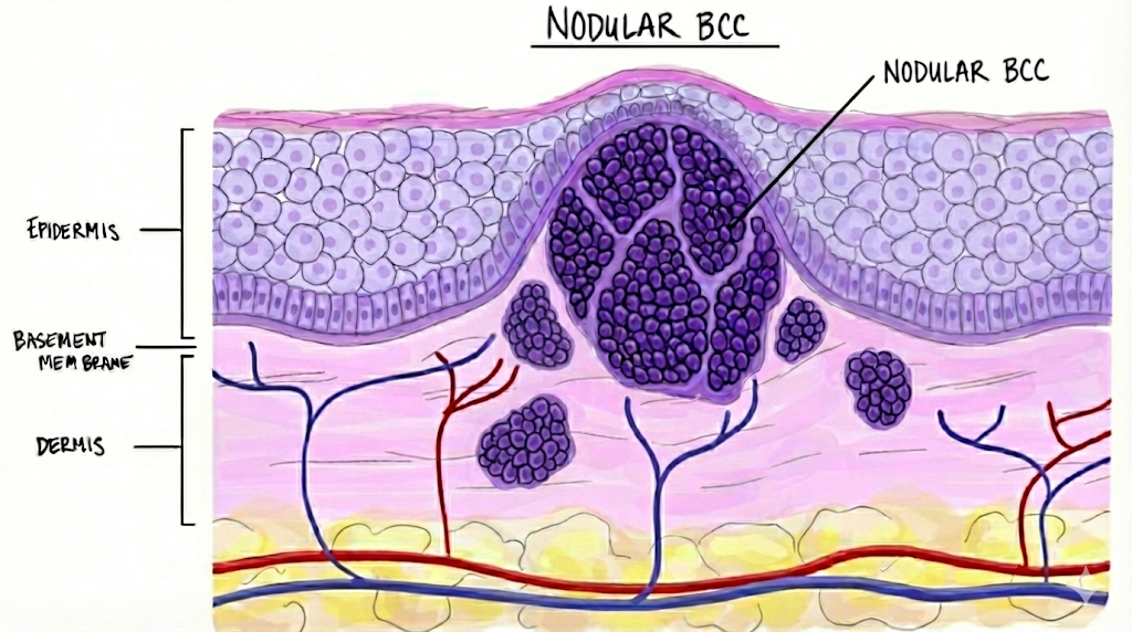
Step 1 — Erbium laser ablation
Complete destruction of any visible lesion with extremely precise laser ablation. After ablation, microscopic foci of cancer may still exist deeper in the dermis — these are addressed in the next steps.
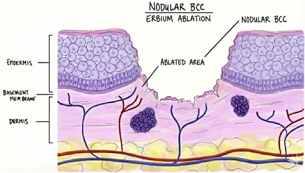
Step 2 — Fractional laser pre-treatment to enhance absorption
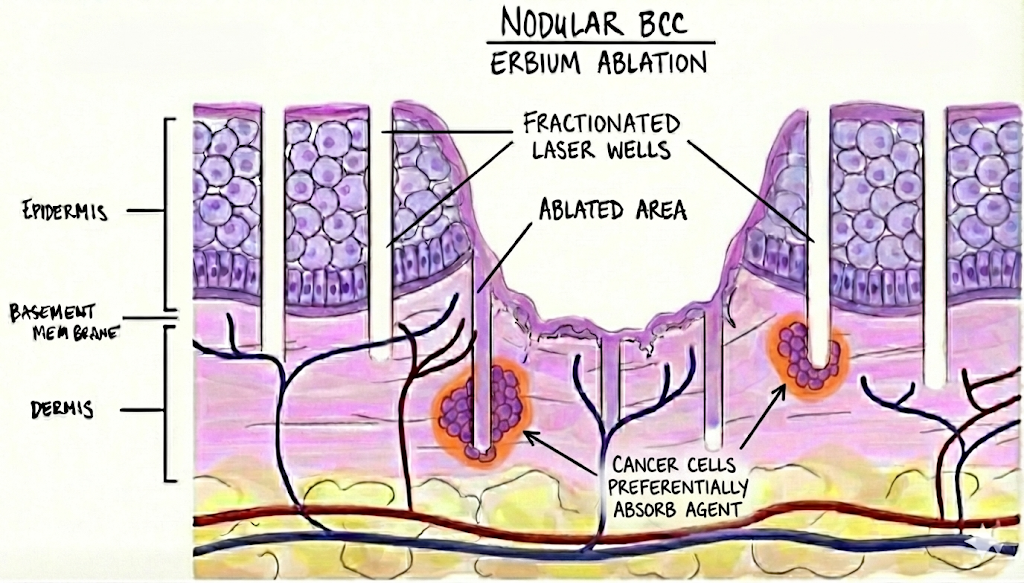
A fractional ablative laser is then used to create microscopic channels into the skin. These “wells” help the medicated cream in the next step penetrate more effectively into the deeper skin and adnexal structures, and also destroy some abnormal cells directly.
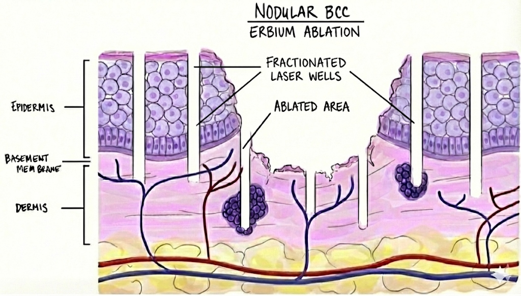
Step 3 — Photosensitising cream (MAL / Metvix)
A photosensitising cream — methyl aminolevulinate (MAL, Metvix) — is applied to the treatment area and absorbed by abnormal cells over a 3-hour incubation.
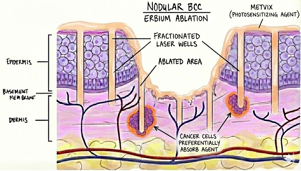
Metvix is a pro-drug. After application, it enters skin cells and is converted through the body’s normal haem (red-blood-pigment) pathway into a natural light-sensitive molecule, protoporphyrin IX (PpIX).
Cancer cells have a more active and “imbalanced” haem pathway than healthy skin. This makes them:
- Take up more of the drug (MAL).
- Make more of the upstream haem-pathway enzymes — converting more of the drug into PpIX.
- Have less of the final enzyme (ferrochelatase) that converts PpIX into safe molecules the light won’t activate.
As a result, PpIX builds up to much higher levels in actinic keratoses and non-melanoma skin cancers than in the surrounding normal skin.
Step 4 — Red light activation
The excess MAL is removed from the skin.
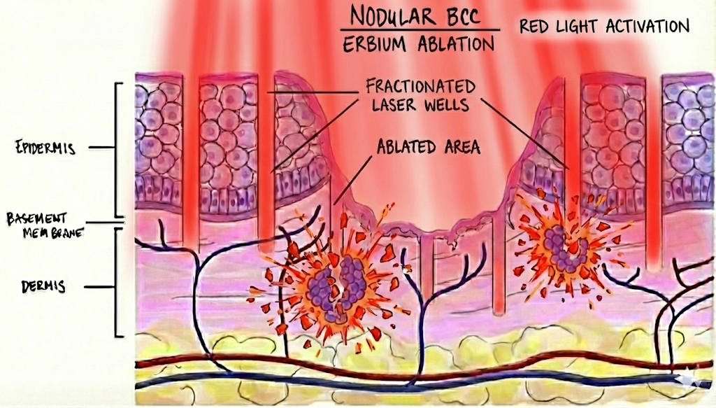
We then shine the red treatment light onto the skin — “red light activation”. This wavelength is designed to specifically interact with PpIX, generating reactive oxygen species (ROS) that preferentially damage and destroy cancerous and precancerous cells, while normal skin (with much lower PpIX levels) is relatively spared.
Treatment comparison: at-a-glance
| Feature | LA-PDT (laser-assisted) | Surgery | Efudix (5-fluorouracil) | Aldara (imiquimod) |
|---|---|---|---|---|
| Indicated for | sBCC, SCCis, nBCC* 8–13 | sBCC, SCCis, nBCC* | SCCis 3 | sBCC 2 |
| Effectiveness | ~94–100% 8–12 | ~97% 20 | 70% 18 | ~80% (non-facial sBCC) 18,19 |
| Delivery | In-clinic, doctor-led | In-clinic | At-home, self-applied 3 | At-home, self-applied 2 |
| Typical course | 1–2 sessions | 1 session | Twice daily for 6 weeks | 5 days a week for 6 weeks |
| Mechanism | Laser ablation + light-activated cell death | Surgical excision | Topical chemotherapy 3 | Immune-system stimulant 2 |
| Visible reaction | Red, weeping area | Scarring | Red, weeping area | Red, weeping area |
| Systemic (whole-body) side effects | None | None | 5–10% can develop flu-like illness, abdominal cramps, persistent vomiting, bloody diarrhoea or alopecia; rare bone-marrow suppression; very rarely life-threatening 14,15,17 | 5–10% can develop flu-like illness, headaches, dizziness, insomnia or diarrhoea 16,17 |
| Discontinuation before treatment complete due to severe side effects | None | None | 5% 17 | 3% 17 |
| Downtime | Short (days) | Weeks – 3 months (dependent on area and size) | Prolonged (weeks–two months) | Prolonged (weeks–two months) |
| Cosmetic outcome | Excellent 8–12 | Good (dependent on area and size) | Good (risk of pigment changes) | Good (risk of pigment changes) |
*nBCC indication for LA-PDT may be considered off-label depending on clinical selection. The pivotal imiquimod (Aldara) superficial-BCC trials excluded facial lesions, so face-specific cure rates are not well established. 19
The clinical evidence — detailed efficacy of LA-PDT by cancer type
The “micro-channelling” and precise ablation combined with PDT used in this pathway allow significantly higher success rates than traditional methods: 8–12
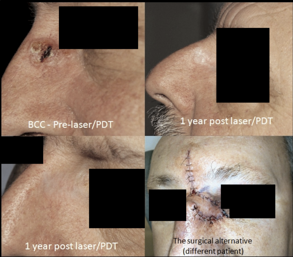
- Superficial BCC (sBCC): Near-perfect outcomes, with studies showing up to 100% clearance 9 and long-term recurrence-free rates of 97.1%. 8
- SCC in situ (Bowen’s disease): Highly effective, with 93.8% clearance. 10
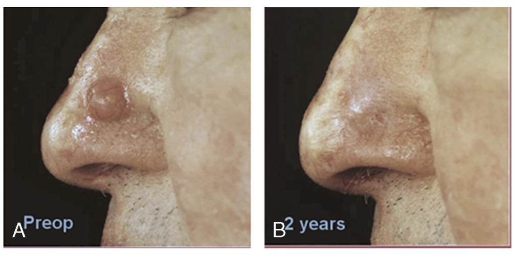
- Nodular BCC (nBCC): Exceptionally high success for a non-surgical method, reaching a 98.97% cure rate when laser ablation is combined with PDT. 11
Outcomes
Excellent cosmetic results after healing
LA-PDT is gentle on surrounding healthy skin. Visible scarring or permanent pigmentation change is minimal. Many patients are highly satisfied with the appearance of their skin after healing. 11
Book your assessment
Suitability for LA-PDT is determined by expert assessment of your lesion’s type, depth and location. Book a consultation to discuss your options.
Book a 20-minute appointment with Dr ChrisRelated pages
What to expect
-
Confirmation of diagnosis
Clinical examination and dermoscopy, followed by biopsy and histology where there is any diagnostic uncertainty. Treatment is only undertaken once the lesion type and depth are confirmed.
-
Step 1 — Erbium laser ablation
The visible cancer is precisely ablated with an erbium laser to remove the bulk of the tumour. Any remaining microscopic disease in the dermis is addressed in subsequent steps.
-
Step 2 — Fractional ablative laser pre-treatment
A fractional ablative laser creates microscopic "wells" in the skin. These channels enhance penetration of the photosensitising cream into deeper skin and adnexal structures and also directly destroy some abnormal cells.
-
Step 3 — Application of methyl aminolevulinate (MAL / Metvix)
A photosensitising cream — methyl aminolevulinate (MAL, Metvix) — is applied to the treatment area. Over a 3-hour incubation, cancer cells preferentially take up the drug and convert it into protoporphyrin IX (PpIX), a light-sensitive molecule.
-
Step 4 — Red LED light activation
Excess cream is wiped away. Red treatment light is then shone onto the skin. It interacts specifically with PpIX inside cancerous and precancerous cells, generating reactive oxygen species that selectively destroy those cells while sparing surrounding healthy skin.
Results timeline
- Immediately after treatment Treated area is red, swollen and tender. A weeping or crusted reaction develops over the next 24–48 hours.
- 5–7 days Crust separates, skin re-epithelialises. Most patients heal within a few days — significantly faster than topical creams or surgery.
- 6–8 weeks Clinical review with dermoscopy to confirm clearance. Any residual lesion is identified and a further treatment or biopsy planned.
- 12 months and ongoing Long-term review for recurrence. Published series show recurrence-free rates of around 97% for sBCC at long-term follow-up.
Ideal candidate
- Patients with biopsy-confirmed superficial basal cell carcinoma (sBCC), SCC in situ (Bowen's disease) or selected nodular BCC.
- Patients who want a non-surgical, in-clinic alternative to Efudix (5-fluorouracil) or Aldara (imiquimod) creams.
- Patients prioritising cosmetic outcome on cosmetically sensitive sites such as the face.
- Patients unable or unwilling to commit to 4–6 weeks of daily at-home cream application.
- Patients without active infection or photosensitivity disorders affecting the treatment area.
Frequently asked questions
-
How effective is laser-assisted PDT for non-melanoma skin cancer?
Published series report up to 100% clearance for selected superficial BCC, ~93.8% clearance for SCC in situ (Bowen's disease), and up to ~98.97% cure for nodular BCC when laser ablation is combined with PDT. -
Is laser-assisted PDT painful?
The laser ablation steps are performed with topical and, where needed, infiltrative local anaesthetic. The red LED light activation phase can cause a stinging or burning sensation, which we minimise with cooling and short pauses. -
How does laser-assisted PDT compare to Efudix (5-fluorouracil) or Aldara (imiquimod)?
Laser-assisted PDT is completed in a single in-clinic session with healing usually within 5–7 days. Efudix (5-fluorouracil) and Aldara (imiquimod) require 4–6 weeks of at-home application and produce weeks of inflammation. Effectiveness is also typically higher with laser-assisted PDT. -
When is surgery still the better option?
Surgery is preferred for higher-risk lesions, deeper or aggressive subtypes, recurrent lesions, where absolute margin control is required, or in selected anatomic sites where laser-assisted PDT is less reliable. -
Is laser-assisted PDT for nodular BCC always appropriate?
Use of laser-assisted PDT for nodular BCC is considered off-label and depends on clinical selection. We discuss the evidence and trade-offs versus surgery in consultation.
References
- Shumack SP. Non-surgical treatments for skin cancer. Australian Prescriber (2011).
- DermNet NZ. Imiquimod.
- DermNet NZ. Fluorouracil (5-FU) cream.
- DermNet NZ. Basal cell carcinoma.
- Griffin LL, Lear JT. Photodynamic therapy and non-melanoma skin cancer. Cancers (Basel) (2016).
- Choi SH, et al. Efficacy of ablative fractional laser-assisted photodynamic therapy for the treatment of actinic cheilitis — 12-month results of a prospective, randomized, comparative trial. Br J Dermatol (2015).
- Steeb T, et al. Laser-assisted photodynamic therapy — mechanistic and comparative context. J Am Acad Dermatol (2019).
- Shokrollahi K, et al. Combined carbon dioxide laser with photodynamic therapy for nodular and superficial basal cell carcinoma. Ann Plast Surg (2014).
- Genouw E, et al. Laser-assisted photodynamic therapy for superficial basal cell carcinoma and Bowen disease — a randomized intrapatient comparison between continuous and fractional ablative CO2 laser. J Eur Acad Dermatol Venereol (2018).
- Ko DY, et al. A randomized trial comparing methyl aminolaevulinate PDT with and without Er:YAG ablative fractional laser in Asian patients with lower extremity Bowen disease — 12-month follow-up. Br J Dermatol (2014).
- Smucler R, Vlk M. Combination of Er:YAG laser and photodynamic therapy in the treatment of nodular basal cell carcinoma. Lasers Surg Med (2008).
- Lippert J, et al. Fractional carbon dioxide laser improves nodular basal cell carcinoma treatment with photodynamic therapy. Dermatol Surg (2013).
- DermNet NZ. Photodynamic therapy.
- Kishi P, Price CJ. Life-Threatening Reaction with Topical 5-Fluorouracil. Drug Safety Case Reports (2018).
- Cohen PR. Topical 5-fluorouracil 5% cream associated with severe neutropenia — case and review of systemic reactions. Dermatol Online J (2018).
- Pasadyn SR, et al. Topical Imiquimod Induces Severe Weakness and Myalgias After Three Applications — A Case Report. J Clin Aesthet Dermatol (2019).
- Love WE, Bernhard JD, Bordeaux JS. Topical imiquimod or fluorouracil therapy for basal and squamous cell carcinoma — a systematic review. Arch Dermatol (2009).
- Jansen MHE, et al. Five-year results of a randomized controlled trial comparing effectiveness of photodynamic therapy, topical imiquimod, and topical 5-fluorouracil in patients with superficial basal cell carcinoma. J Invest Dermatol (2018).
- Raasch B. Management of superficial basal cell carcinoma — focus on imiquimod. Clin Cosmet Investig Dermatol (2009). Pivotal imiquimod sBCC efficacy (~78–82%) applies to lesions not on the face or neck.
- Thomson J, et al. Interventions for basal cell carcinoma of the skin. Cochrane Database of Systematic Reviews (2020).
Related
Related conditions
Medically reviewed by Dr Christopher Irwin, MBChB, FRACGP, MMed (Skin Cancer), FACAM, MSCCA · Last reviewed 2026-06-06 · Editorial policy