
- Thin invasive melanoma (Stage I) has a Breslow thickness under 1 mm — roughly the thickness of a grain of sand.
- Risk of metastasis is low — under 5% for melanomas thinner than 0.8 mm, around 5–12% for those between 0.8 and 1.0 mm.
- Treatment is wide local excision with a 1 cm margin of normal-looking surrounding skin.
- Sentinel lymph node biopsy (SLNB) is reserved for Breslow 0.7–1 mm with high-risk features (ulceration, mitotic rate >1, Clark IV/V, lymphovascular invasion); overall complication rate ~11.3%.
- Routine CT/PET scans are not recommended — yield is extremely low and the risks outweigh the benefit.
- Five-year survival is 95–99% with proper treatment and structured follow-up.
- Skin checks every 3–6 months for the first two years, then 6–12 monthly long-term; risk of a new primary melanoma is elevated lifelong.
- Care provided across two Melbourne clinics — Ivanhoe and Diamond Creek.
Quick definition
A thin invasive melanoma (also called thinly invasive melanoma or early Stage I melanoma) is an invasive melanoma with a Breslow thickness less than 1 mm — about the thickness of a grain of sand. The melanoma has crossed the basement membrane into the dermis, but only slightly. Risk of spread is under 5% for the thinnest tumours and around 5–12% for those approaching 1 mm. Standard treatment is wide local excision with a 1 cm margin, and five-year survival is 95–99%. Routine CT/PET scans and lymph node biopsy are usually not required.
Being diagnosed with melanoma can be scary, but the good news is that the type of melanoma you have, even though it is invasive, is still early-stage and will most likely be completely cured by simple surgical removal with margins. This handout explains thinly invasive melanoma — and why it usually does not require additional tests like lymph node biopsies or body scans. We’ll also discuss what to expect next and how these early melanomas are managed in a patient-friendly way.
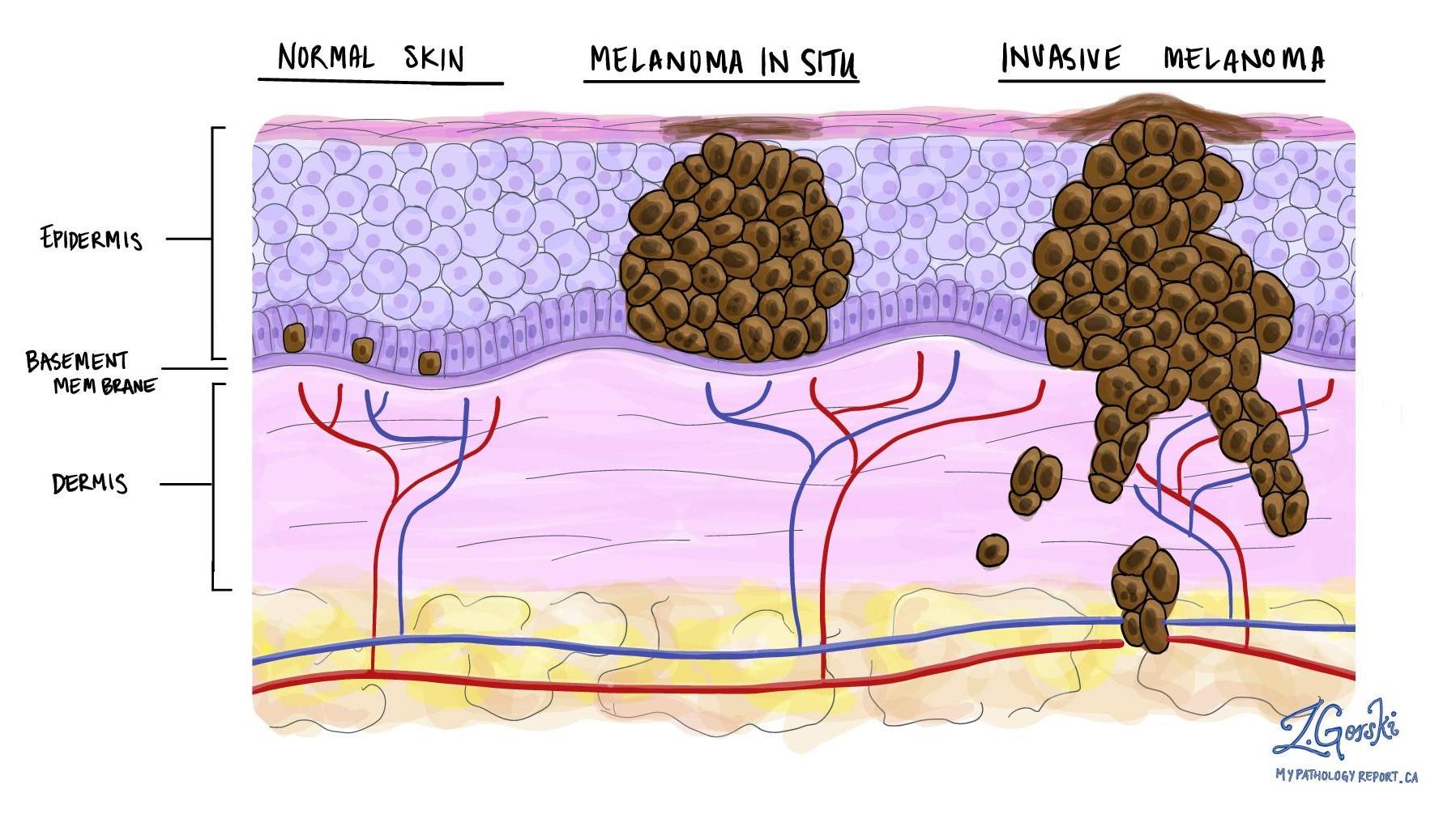
If your report says you have an invasive melanoma with a Breslow thickness less than 1 mm, this is often called a thinly invasive melanoma and corresponds to an early Stage I melanoma. “Invasive” simply means the melanoma did grow deeper than the epidermis into the second layer of skin (dermis) — breaking through the strong canvas layer called the basement membrane or “dermo-epidermal junction” — but in this case only slightly.
Thinly invasive melanoma is more serious than in situ melanoma because there is a low chance of spread through the body. How serious an invasive melanoma is depends on several variables. The primary “badness” marker is the Breslow thickness.
Understanding Breslow Thickness
Breslow thickness is a measurement in millimetres from the top of the skin (technically the granular layer of the epidermis) to the deepest point of invasion by the melanoma. Let’s put this into perspective: 1 mm is the same thickness as a grain of sand. If you have a melanoma less thick than a grain of sand, the most likely outcome if you are treated properly is that everything will be completely fine.
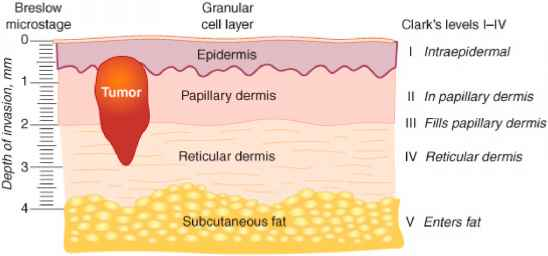
The reason we care most about the Breslow thickness is that it is the main marker for how likely a melanoma has spread to regional lymph nodes (like in the armpits or groin) — or further.
In general, the risks for spread for a thinly invasive melanoma are really low — less than 5%.
Other risk factors on the histology report
Other features on the histology report that make spread more likely are:
- Ulceration
- Mitotic rate >1
- Clark level IV or V (extending into the reticular dermis or into fat — though this is very unlikely in melanoma under 1 mm thick)
- Lymphovascular invasion (LVI)
Histology reports are very complicated and have a whole bunch of other words on them. If you have any questions about the specific words feel free to ask during your consult. In the vast majority of cases, anything other than the Breslow thickness and the risk factors above does not really alter management.
Do I Need Further Scans or Tests?
In general, no. For most patients with thinly invasive melanoma, the risks of the scans or tests outweigh the benefit they provide.
Current guidelines in Australia state that if a melanoma has a Breslow thickness of 0.7 mm–1 mm AND has any of the risk factors above (ulceration, mitotic rate >1, Clark level IV or V or lymphovascular invasion), we discuss the role of a Sentinel Lymph Node Biopsy (SLNB).
In basic terms a SLNB is a procedure to see whether the closest lymph node contains any melanoma cells that have spread from the skin. It requires a general anaesthetic (being put to sleep) and has some significant risks associated with it. The overall risk of a complication from SLNB is around 11.3%. While it is important to consider this procedure if your risk of spread is >5%, we don’t recommend this procedure for most low-risk thinly invasive melanomas, as we are more likely to harm you than help you with the procedure.
Key Facts About Thinly Invasive Melanomas
- Low risk of spread. Thin melanomas have a very low risk of metastasis — slightly higher than in situ. The risk of cancer cells having spread to lymph nodes or beyond is under 5% for melanomas thinner than 0.8 mm, and in the range of about 5–12% for those between 0.8 and 1.0 mm.
- Surgery is the main treatment. Just like melanoma in situ, the first step is a wide local excision surgery to remove the melanoma with a safety margin of normal skin. Typically, a margin of 1 cm of normal skin is taken for melanomas up to 1 mm thick (per guidelines) to ensure clear margins.
- Sentinel lymph node biopsy usually not needed. Because the chance of spread is so low, additional lymph node testing isn’t usually recommended for these thin melanomas. Medical guidelines do not recommend a sentinel lymph node biopsy for most thinly invasive melanomas.
- No routine scans needed. Full-body imaging scans (CT, PET, etc.) are not done for a thin melanoma that shows no signs of spread. The chance of any scan finding melanoma in other organs at this early stage is extremely low, so doctors avoid exposing you to unnecessary radiation or causing anxiety from false alarms.
- Excellent prognosis. Survival rates are above 95–99% at 5 years for Stage I melanoma patients.
The vast majority of people in this category remain cancer-free after treatment. You will still need regular skin checks (often every 3–6 months for the first two years) to watch for any new melanomas or changes, but the melanoma you had is very unlikely to come back after proper removal.
Follow-Up and Outlook
Your prognosis is extremely good. Both melanoma in situ and thin invasive melanomas have high cure rates after proper removal — meaning the vast majority of patients remain alive and well, with no recurrence of melanoma. Many experts would say you are likely cured at this point. Here is what to expect moving forward, and some tips for peace of mind.
- Regular skin checks. You will need periodic skin checks and reviews with your skin doctor. Typically, for a thin melanoma, the schedule might be every 3–6 months for the first couple of years, before moving to a long-term 6–12 month review cycle. These visits are to ensure no new melanoma (or other skin cancers) develop and to catch any changes early. Self skin exams at home monthly are also a good habit — your doctor can teach you how to examine your moles and what to look for.
- No routine scans or blood tests. As discussed, you won’t be undergoing regular CT/PET scans or blood work for melanoma markers, because there’s no indication to do so. Try not to be alarmed by the lack of testing — it truly is a positive sign about your prognosis. Of course, if you develop any new symptoms (a new lump in your groin/armpit, unexplained persistent pain, etc.), inform your doctor. They will investigate appropriately. But such scenarios are not expected given how early your melanoma was.
- Scar care. You will have a scar from the surgery. Keep it clean and protected from the sun as it heals. Over time (months to a year) it will fade and soften. Your doctor can advise on great dressings like MEFIX, scar creams or silicone gel sheets if needed. Most patients find the scar becomes a minor cosmetic issue, especially compared to the relief of being cancer-free. The more care you take for your scar in the first 3–6 months, the better the long-term scar will look.
- Sun protection. One of the best ways to prevent future skin cancers is to practice good sun safety. That means using sunscreen (SPF 50) on exposed skin daily and reapplying every 2 hours whenever outside, wearing hats and protective clothing.
- Peace of mind. It is normal to feel anxious after a cancer diagnosis, even one as low-risk as this. Remember that your melanoma was caught at a very early stage. If you find yourself worrying a lot, consider discussing these feelings with your doctor. Sometimes connecting with a counsellor or a support group of melanoma survivors can be helpful. Knowing others have gone through the same thing and are doing fine can be very reassuring.
Frequently asked questions
-
What does "thinly invasive melanoma" mean?
It means an invasive melanoma with a Breslow thickness less than 1 mm — corresponding to an early Stage I melanoma. "Invasive" means the melanoma has grown beyond the epidermis through the basement membrane into the dermis below, but in a thin melanoma only slightly. To put 1 mm in perspective, it is roughly the thickness of a grain of sand. -
What is Breslow thickness and why does it matter?
Breslow thickness is the measurement in millimetres from the top of the skin (the granular layer of the epidermis) down to the deepest melanoma cell. It is the single most important prognostic marker because it best predicts how likely the melanoma is to have spread to regional lymph nodes (armpits, groin) or beyond. Other histology features (ulceration, mitotic rate >1, Clark level IV/V, lymphovascular invasion) refine that estimate. -
Do I need a sentinel lymph node biopsy (SLNB)?
Usually no. Current Australian guidelines say SLNB is discussed when Breslow thickness is 0.7–1 mm AND there is at least one high-risk feature (ulceration, mitotic rate >1, Clark IV/V, lymphovascular invasion). SLNB requires a general anaesthetic and carries an overall complication rate of around 11.3%, so for most low-risk thin melanomas it is more likely to harm than help. -
Do I need CT, PET or other scans?
No. Routine full-body imaging is not recommended for a thin melanoma showing no signs of spread. The chance of a scan finding melanoma in another organ at this early stage is extremely low, and the radiation, cost and risk of false alarms outweigh any benefit. -
What is the prognosis (five-year survival)?
Excellent — survival is 95–99% at 5 years for Stage I melanoma patients. The risk of metastasis is under 5% for melanomas thinner than 0.8 mm and around 5–12% for those between 0.8 and 1.0 mm. -
How is thin invasive melanoma treated?
Wide local excision with a 1 cm margin of normal-looking surrounding skin, for melanomas up to 1 mm thick (per guidelines). The specimen is sent to a pathologist to confirm clear margins. Surgery is the main treatment; additional procedures are reserved for selected high-risk features. -
How often will I need skin checks afterwards?
Typically every 3–6 months for the first two years (when the risk of a new primary is highest), then extending to every 6–12 months long-term. Monthly self-skin-exams at home are also recommended — your doctor can teach you what to look for. The melanoma you had is very unlikely to come back after proper removal; checks are mainly to catch any new primaries early.
References
- Clinical practice guidelines for the diagnosis and management of melanoma. Cancer Council Australia & Melanoma Institute Australia.
- Cutaneous melanoma. Lancet. 2023. (Long GV, Swetter SM, Menzies AM, Gershenwald JE, Scolyer RA.)DOI: 10.1016/S0140-6736(23)00821-8
- NCCN Guidelines Insights — Melanoma (Cutaneous), Version 2.2024. J Natl Compr Canc Netw. 2024.DOI: 10.6004/jnccn.2024.0036
Related
Medically reviewed by Dr Christopher Irwin, MBChB, FRACGP, MMed (Skin Cancer), FACAM, MSCCA · Last reviewed 2026-06-06 · Editorial policy