
- SCC in situ is the earliest form of SCC — confined to the epidermis, has not crossed the basement membrane.
- Also known as Bowen's disease or intraepidermal carcinoma (IEC) — the same condition under different names.
- Zero risk of having already spread because the blood and lymph vessels cancer uses to spread exist only in the dermis below.
- Looks like a persistent red or pink scaly patch — often mistaken for eczema or psoriasis.
- One step beyond actinic keratosis — full-thickness epidermal abnormality vs lower-epidermal in AK.
- Can progress to invasive SCC over months to years if untreated — treatment is recommended.
- Multiple treatment options — topical 5-fluorouracil (Efudix, ~70%), laser-assisted PDT (>90–95%), curettage or surgical excision.
- No CT/PET scans, blood tests or lymph node biopsy required.
- Care provided across two Melbourne clinics — Ivanhoe and Diamond Creek.
Quick definition
SCC in situ — also known as Bowen’s disease or intraepidermal carcinoma (IEC) — is the earliest form of squamous cell carcinoma. The cancer cells are confined to the epidermis (the top layer of skin) and have not crossed the basement membrane into the dermis. Because the vessels cancer uses to spread exist only in the dermis below, SCC in situ has zero risk of having spread. It usually looks like a persistent red or pink scaly patch and is often mistaken for eczema or psoriasis. Treatments: topical 5-fluorouracil (Efudix) ~70%, laser-assisted photodynamic therapy >90–95%, curettage or surgical excision. Outstanding prognosis.
Squamous cell carcinoma (SCC) is the second most common type of skin cancer (after BCC). SCC is divided into two categories based on how deep it goes: in situ (only in the top layer) and invasive (growing into deeper layers). This page covers SCC in situ.
What Is SCC in Situ?
SCC in situ means the cancer cells are confined to the top layer of the skin (the epidermis) and have not invaded deeper layers. It is also known as “Bowen’s disease” or “Intraepidermal carcinoma” (IEC). You might just see a persistent red, scaly patch that doesn’t heal — it can look like a rash or eczema patch.
In situ SCC is basically the earliest form of SCC (think of it as “stage 0” skin cancer). It grows very slowly over months to years and stays where it started. Because it hasn’t grown inward, it cannot spread (metastasize) to lymph nodes or other organs at this stage.
Why the basement membrane matters
The skin contains two basic parts. The epidermis is the top layer of skin and the bottom layer of skin is called the dermis. The epidermis and dermis are separated by a very tough canvas-like layer called the basement membrane (or “dermo-epidermal junction”). If a cancer has not yet learnt how to get through this tough canvas layer, that means it has no way of travelling to other parts of the body. This is because all of the highways that cancers use (veins, arteries and lymph vessels) are all only in the dermis. There are no highways in the epidermis. This is great because it means if we treat it properly, the cancer can essentially be cured with no risk of metastasis.
The main concern is that, if left untreated, it could eventually turn into invasive SCC.
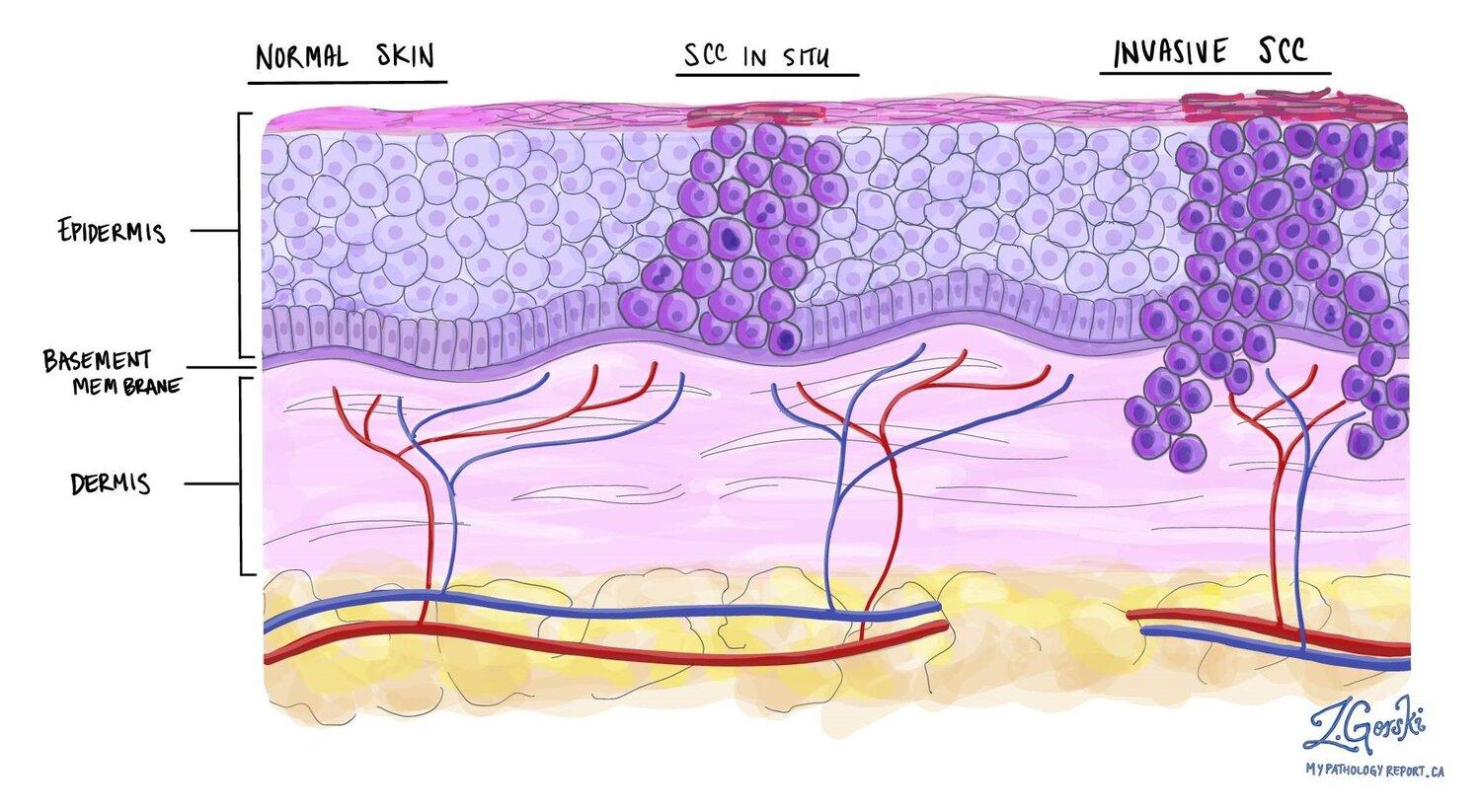
Risk of Spread
By definition, in situ SCC has zero risk of having already spread elsewhere, since it hasn’t grown into the dermis. However, if not treated, in situ lesions can become invasive over time. Because of this chance of progression, doctors recommend treating Bowen’s disease so it doesn’t get the chance to turn into an invasive SCC. The good news is that once it’s treated, it’s essentially cured and no longer poses a threat.
Are Lymph Node Biopsies or Scans Needed?
No — not for in situ SCC. Because it has not invaded the skin, there is virtually zero chance of spread to lymph nodes or elsewhere at this stage. Therefore, no CT scans or lymph node biopsies are indicated for isolated SCC in situ. The focus is just on treating the patch on the skin.
Outlook
The outlook for SCC in situ is excellent. Once treated, it is usually cured outright. Bowen’s disease itself is not life-threatening. The main goal is preventing progression to invasive SCC, and treatment achieves that in the vast majority of cases. We do recommend regular check-ups because having Bowen’s disease can be a sign of sun damage, and you might develop other skin cancers or another patch in the future.
Symptoms
- A persistent red, pink or brown scaly patch that does not heal
- Often slightly raised at the edge, with a flat, well-defined border
- May crust, fissure, or weep occasionally
- Painless or mildly itchy — easy to mistake for eczema or psoriasis
- Slowly enlarges over months to years
- Most common on sun-exposed skin (lower legs, face, scalp, hands) but can occur anywhere
Causes & contributors
- Cumulative ultraviolet (UV) exposure from sunlight and solariums
- Fair skin, light eyes, easy burning
- Older age (although younger patients with heavy sun damage are common in Australia)
- A field of multiple actinic keratoses (sun-damage pre-cancer)
- Immunosuppression — transplant recipients have substantially higher SCC risk
- HPV infection in selected sites (eg. genital, periungual)
Diagnosis
Diagnosis is made on clinical examination and dermatoscopy and confirmed on histology after a small biopsy. The pathology report names the lesion as "SCC in situ," "Bowen's disease," or "intraepidermal carcinoma" — all three terms describe the same condition. No scans, blood tests or lymph node biopsy are needed because the cancer has not crossed the basement membrane and has no route to spread.
Treatment options
Topical 5-fluorouracil (Efudix) →
Prescription cream applied to the patch over six weeks. Kills cancerous cells. The area becomes red and crusty during treatment — this is normal as abnormal cells die off. Around 70% effective.
Laser-assisted photodynamic therapy (LA-PDT) →
One of the most common treatments we perform for SCC in situ. Initial laser ablation, then a fractional laser creates "wells" for cream uptake, then a photosensitising cream is applied and activated by red LED light. Short downtime and >90–95% success — excellent cosmetic outcomes.
Curettage and electrodessication
The skin is numbed and the abnormal cells are scraped off, then the base is cauterised. Quick procedure but leaves more scarring than topical or PDT options. A pragmatic choice for patients with multiple lesions who want them resolved quickly.
Surgical excision
The patch is cut out under local anaesthetic and stitched closed. Good cure rate but more scarring than non-surgical options for surface-only lesions. Reserved for selected cases.
When to see a doctor
See a doctor if you have a persistent red or pink scaly patch on sun-exposed skin (especially the lower legs, face, scalp or hands) that has been present for weeks to months and is not healing — particularly if you have treated it as eczema or psoriasis without improvement. A small biopsy can confirm the diagnosis and several effective treatments are available.
Frequently asked questions
-
Is SCC in situ cancer?
Yes, technically — but it is the earliest possible form of squamous cell carcinoma (Stage 0). The cancer cells are confined to the very top layer of skin (the epidermis) and have not crossed the basement membrane into the dermis. Because the blood and lymph vessels cancer uses to spread exist only in the dermis, SCC in situ has zero risk of having spread. It still needs treatment to prevent progression to invasive SCC. -
Is SCC in situ the same as Bowen's disease?
Yes — they are the same condition under different names. "SCC in situ," "Bowen's disease," and "intraepidermal carcinoma" (IEC) all refer to a squamous cell carcinoma confined to the epidermis. Different doctors and pathology reports use different terms — they all mean the same thing. -
What does SCC in situ look like?
A persistent red, pink or brown scaly patch that does not heal. The patch is often slightly raised at the edges with a well-defined border, may crust or fissure occasionally, and is usually painless or only mildly itchy. It is often mistaken for eczema or psoriasis because of the persistent scale. Most commonly found on sun-exposed skin (lower legs, face, scalp, hands). -
What is the difference between SCC in situ and actinic keratosis?
Actinic keratosis (AK) is a pre-cancer — the abnormal cells occupy only the lower part of the epidermis. SCC in situ has progressed further — abnormal cells now occupy the full thickness of the epidermis. Both are confined above the basement membrane and have no metastatic risk, but SCC in situ is one step closer to invasive SCC. AKs that are persistent, thick, or tender are more likely to progress and should be treated. -
Does SCC in situ spread to lymph nodes or other organs?
No. By definition, SCC in situ has not crossed the basement membrane into the dermis, and the vessels cancer uses to spread only exist in the dermis. Zero risk of metastasis as an in-situ lesion. No CT scans, PET scans, blood tests or lymph node biopsies are needed. -
Will SCC in situ turn into invasive SCC if I don't treat it?
It can. Untreated, SCC in situ can progress to invasive SCC over time — though the rate is slow (months to years). Estimates vary, but a meaningful minority of untreated lesions eventually become invasive. This is why treatment is recommended even though the in-situ lesion itself poses no metastatic risk. -
Can SCC in situ be treated with cream instead of surgery?
Yes. Topical 5-fluorouracil (Efudix) applied over six weeks is one of the standard treatments — around 70% effective. Laser-assisted photodynamic therapy (LA-PDT) is an in-clinic alternative with >90–95% success and short downtime. Surgical excision is reserved for selected cases where non-surgical options are not suitable. -
How effective is laser-assisted PDT for SCC in situ?
Highly effective — >90–95% success in suitable lesions. The treatment combines laser ablation, fractional laser "wells" to enhance cream uptake, a photosensitising cream, and red-LED light activation. Short downtime (usually a few days), excellent cosmetic outcomes — one of the most common treatments we use for SCC in situ.
References
- Clinical practice guidelines for keratinocyte cancer. Cancer Council Australia. (NHMRC-approved 2019.)
- Guidelines of care for the management of cutaneous squamous cell carcinoma. J Am Acad Dermatol. 2018. (American Academy of Dermatology.)DOI: 10.1016/j.jaad.2017.10.007
- Cutaneous squamous cell carcinoma — an updated review. Cancers (Basel). 2024.DOI: 10.3390/cancers16101800
Related
Related conditions
Medically reviewed by Dr Christopher Irwin, MBChB, FRACGP, MMed (Skin Cancer), FACAM, MSCCA · Last reviewed 2026-06-06 · Editorial policy